Electronic & Chemical & Magnetic Characterization
Magnetic characterisation View all
MFDC Magnetic/ferroelectric/dielectric char.

We provide modular experimental stations to study the evolution of electric and magnetic dipole orders, and their degree of coupling, which is an identifying feature of novel magneto-electric systems. Features for designing multifunctional devices, such as magneto-electric sensors or high-capacity four-state logic memories, can be assessed.
EPR Electron Paramagnetic Resonance

EPR technique allows the detection and study of transient and stable paramagnetic species such as free radicals, over a very wide range of temperatures.
SQUID Superconducting Quantum Interference Device

SQUID offers high resolving power in deciphering static and dynamic phenomena in low-moment, dilute magnetic systems, by measuring moment versus applied magnetic field or moment versus temperature. It also allows precise determination of the Curie, Néel or blocking temperatures of ferromagnetic, antiferromagnetic or superparamagnetic nanoparticles.
Magnetometry Magnetometry

We provide an automated MagLab-EXA multi-measurement system for nanomaterials characterization. A set of analytical probes (AC/DC magnetometry, magneto-transport, Hall effect etc) is available to indentify technologically useful quantum mechanical phenomena to design materials with optimal device operational capabilities.
MT Magneto-transport

We offer magneto-transport characterizations of semiconductor and metallic samples at temperatures down to liquid He and fields up to 7T. Density and mobility of carriers are measured through Hall effect, as well as quantum transport phenomena in 2D systems at low T. Carrier populations can be tuned by bias voltages and external illumination.
MOKE Magneto-Optic Kerr Effect
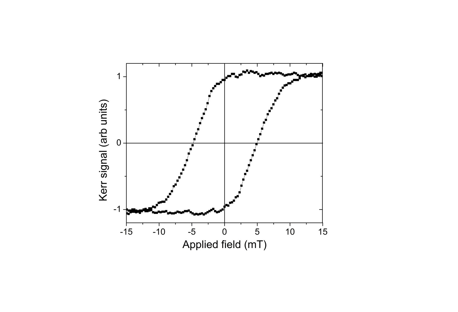
By MOKE the magnetic properties (e.g. coercivity, squareness ratio) of single micro-structures can be evaluated: no quantitative measure of the magnetization is provided, but from the shape of the hysteresis loop, both static and dynamic magnetization reversal processes can be investigated in detail.
Spectro-microscopy View all
AES/SAM Scanning Auger Microscopy

Auger Electron Spectroscopy (AES) and SAM are electron beam techniques allowing the analysis of the surface elemental composition of materials. By pointing or scanning the electron beam, localized information or spatial elemental distributions can be obtained. Auger depth profiling allows measuring elemental concentration profiles.
XPEEM/kPEEM/SPEM PhotoEmission Microscopy

Photoemission microscopy combines XPS with high spatial resolution, achieved by a small spot X-ray beam or by a magnifying electron optical column. Surface-sensitive images are provided for investigating processes occurring at morphologically/chemically complex solid surfaces including chemical reactions and mass transport processes.
SPELEEM spectroscopic photoemission and low energy electron microscope
The Spectroscopic PhotoEmission and Low Energy Electron Microscope (SPELEEM) installed at the MAXPEEM beamline offers unique possibilities for extracting simultaneously elemental, chemical, magnetic and electronic information at spatial resolutions down to the single digit nanometer range to users from a wide area of research fields.
HSI Hyperspectral imaging

Hyperspectral imaging is an advanced hybrid technique that incorporates simultaneously two techniques, imaging and spectroscopy. It finds applications on the characterization of materials with spatial variability like painted surfaces, or materials that have suffered stresses which alter their chemical composition.
MSI Multispectral Imaging

Multispectral imaging is a hybrid technique that allows the simultaneous acquisition of both images and reflection spectra. It is very useful to characterize materials with spatial variability like painted surfaces, or materials that have suffered stresses which alter their chemical composition.
Chemical analysis View all
HPLC HPLC with CAD, UV and Fluorescence detectors(Chemical composition analysis)
HPLC is analytical technique to separate, identify, and quantify molecules in a mixture. Dissolved components are pumped into a packed column where they are separated from each other due to their different degrees of interaction with the packing material. HPLC-UV/MS can be used for the detection and quantification of organic contaminants in water.
AT Automated Titration
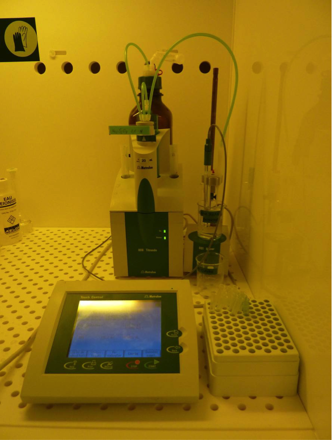
Titration is an analytical technique that allows the quantitative determination of a specific substance (analyte) dissolved in a sample. This technique is based on a complete chemical reaction between the analyte and a reagent (titrant) of known concentration, added to the sample.
ICP-MS ICP Mass spectroscopy (ICP-MS) (single particle analysis, trace element analysis)

ICP-MS allows measuring elements at trace levels. Dissolved sample is introduced into an argon plasma where the molecules are dissociates and further ionized. Singly-charged ions thus formed are directed into the mass spectrometer. A special application of ICP-MS (spICP-MS) allows measuring size and number concentration of a particle suspension.
MOS Mossbauer spectroscopy
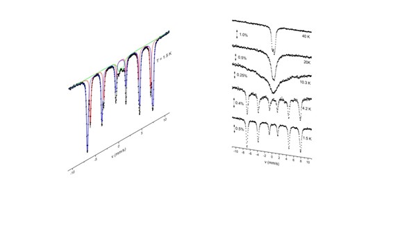
Mössbauer spectroscopy is used for the characterization of electronic and magnetic properties of materials containing specific isotopes such as 57Fe, including alloys and oxides, magnetic nanoparticles for magnetic storage and biomedical applications, synthetic and natural minerals and ores, and iron-containing biological systems and thin films.
Luminescence spectroscopy View all
TR-XRF Total Reflection X-ray Fluorescence Spectrometer (TR-XRF) (Trace element analysis)

TXRF is able to measure the elemental composition of samples (in general for elements with atomic number >12) prepared as a thin film specimen on a polished sample carrier.
FS Fluorescence Spectroscopy
Fluorescence is a type of luminescence caused by photons exciting a molecule, raising it to an electronic excited state. Fluorescence spectroscopy analyzes fluorescence from a molecule based on its fluorescent properties.
CL Cathodoluminescence
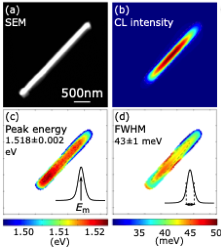
The cathodoluminescence integrates a scanning electron microscope (SEM) and a light microscope into one tool allowing SEM images and electron beam induced-luminescence images to be acquired at the same time. The analysis of the features in the luminescence spectra allows the characterization of the material properties.
FCS Fluorescence Correlation Spectroscopy

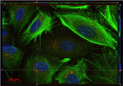
Fluorescence Correlation Spectroscopy (FCS) is a fluorescence based technique that gives information on the diffusion of fluorescent molecules or objects. From the diffusion times, the size of diffusing species can be determined, and the interaction among molecules or nanoparticles can be studied.
ML Microluminescence

The spectrum of the emitted light by the sample, excited through electrical polarization, electronic bombardment, optical illumination or magnetic excitation, provides important information on the electronic structure of the material. Introducing a microscope spatial distribution of the electronic structure can be obtained.
PL PhotoLuminescence

PL is a non-contact, non-destructive method of probing the electronic structure of materials, often used in the context of semiconductor devices to determine the bandgap energy, the composition of heterostructures, the impurity levels, the crystal quality, and to investigate recombination mechanisms.
μ-XRF micro XRF
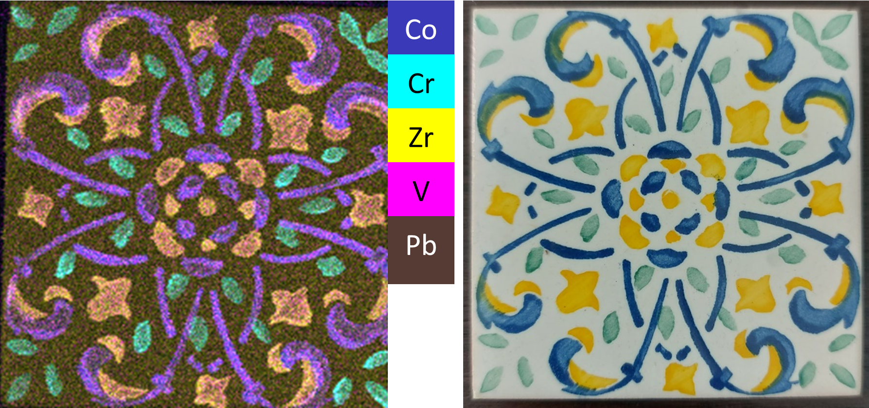
The μ-XRF technique offers non-invasive multi-elemental, qualitative and quantitative analysis of bulk samples and thin films at the micrometre scale (50-500 μm) within a broad analytical (Z>13) and sensitivity (μg/g - %wt.) range. The scanning mode generates elemental distribution maps within ~mm2-cm2 areas with micrometre spatial resolution.
TRLS Time Resolved Luminescence Spectroscopy
P66 beamline, located within PETRA III storage ring, specializes in materials science, enabling precise photoluminescence studies of solids under state-selective excitation via pulsed synchrotron radiation in UHV. It enables recording time-resolved emission and excitation, absorption and reflection spectra across a wide 8-800 K temperature range.
Optical spectroscopy View all
Pump-probe Pump-probe

The pump-probe spectroscopy infrastructure provides in-situ probes of the excited state of the matter, i.e. in the time/frequency domain at the fs-ps scales.
IRS UHV reflection-absorption IR spectroscopy

An infrared spectrum is commonly obtained by passing infrared radiation through a sample and determining what fraction of the incident radiation is absorbed at a particular energy. The peak position in the absorption spectrum corresponds to the frequency of a vibration in the sample molecule.
Ellipsometry Ellipsometry

Ellipsometry is a contact-free, nondestructive method for characterization of the dielectric and optical properties (refractive index, absorption and thickness) of layered nanostructures in the size range of < 1 nm to several μm.
RS Raman spectroscopy

Raman spectroscopy (RS) investigates the vibrational properties of a sample and provides chemical as well as structural information. RS does not require any specific sample preparation, size or condition and may be combined with micron/nano spatial resolution when operated using a confocal microscope/TERS or SNOM configuration.
FTIR Fourier Transform Infra Red Spectroscopy and Spectromicroscopy

IR spectroscopy is based on the absorption of infrared radiation by matter. This leads to energetic transitions in the vibrational state of concrete chemical bonds. Since the frequencies that are absorbed by the molecules are characteristic of their structure, this technique is a powerful tool for qualitative and quantitative molecular analysis.
OS Optical spectroscopy

VUV/UV/Vis/NIR spectroscopy is the measurement of the attenuation of a beam of light after it passes through a sample or after reflection from a sample surface. It is useful to characterize absorption, transmission, and reflectivity of a variety of technologically important materials, such as gases, film, pigments, coatings, windows, and filters.
BLS Brillouin Light Scattering
Brillouin spectroscopy is an empirical spectroscopy technique which allows the determination of elastic moduli of materials. The technique uses inelastic scattering of light when it encounters acoustic phonons in a crystal, a process known as Brillouin scattering, to determine phonon energies and therefore interatomic potentials of a material.
TG TRANSIENT GRATING

The Transient Grating is a pump-probe technique allowing a direct access to the acoustic, magnetic and thermal degrees of freedom in condensed matter. Thanks to various laser sources, it allow to cover a dynamical range from hundreds of femtoseconds to hundreds of microseconds.
Neutron magnetic characterisation View all
DNS** Diffuse Neutron Scattering (temporarily unavailable)

DNS with single-crystal time-of-flight spectroscopy is ideal for the studies of complex spin correlations, and represents a powerful instrument for the soft condensed matter community for the separation of nuclear coherent scattering from often dominating spin incoherent scattering background in a wide-angle diffraction experiment.
M-SANS** Magnetic Small Angle Neutron Scattering (temporarily unavailable)

SANS is used for the analysis of 1-100 nm structures, which cannot be characterised by microscopies or when samples are turbid due to multiple light scattering. In KWS-1 beamline magnetic samples are studied with the full polarisation analysis. In KWS-2 the contrast variation method allows for highlighting of particular components.
MNRefl** Magnetic Neutron Reflectivity (temporarily unavailable)

With MNR we investigate thin magnetic layered structures down to thicknesses between 3–300 Å and lateral structures from nm to µm, gaining insight into magnetic roughness, magnetic domains, lateral structures...The reflection of polarised neutrons allows to determine density, modulus and direction of the magnetisation vector of buried layers.
X-ray/soft-X-ray spectroscopy View all
IXS Inelastic X-ray Scattering
Inelastic X-ray Scattering (IXS) permits to analyze several aspects of materials' dynamics. It includes X-Ray Raman Scattering to measure soft x-ray atomic absorption edges with a bulk sensitivity, Resonant Inelastic X-Ray Scattering to probe magnetic and orbital excitations in strongly correlated electron systems, and Compton scatterring.
DiProi-FEL Coherent Diffraction Imaging @ Free Electron Laser
DiProI end-station is a multi-porpose experimental chamber dedicated to time resolved imaging and scattering experiments taking advantage of FEL radiation provided by the FERMI source. The instrument is particular appealing for provide ultrafast information on electronic and spin dynamics after an external stimolus.
IOS In-operando spectroscopy

Soft X-rays XAS operating at Ambient Pressure allows for in-operando spectroscopic investigation of surfaces and their catalytical properties. This opens interesting access to L and M-edges of most of the transition metals and K-edges of light elements (C, O and N). The high surface sensitivity is achieved by the total electron yield detection.
XAS X-ray Absorption Spectroscopy

XAS is sensitive to the local bonding environment of the atom absorbing the X-rays, providing information on oxidation states, local orbital symmetry, molecular orientation and chemically selective density of states. It is widely used in molecular and condensed matter physics, material science, engineering, chemistry, earth science and biology.
XMCD/XMLD X-ray Magnetic Circular/Linear Dichroism

When X-ray absorption is measured with circularly/linearly polarized x-rays, spin and angular momenta can be determined in ferromagnetic/antiferromagnetic systems, respectively. Dichroic effects arise by the difference between spectra measured with different helicity/polarization orientation of the X-ray photons.
SXES Soft x-ray Spectroscopy with Electrons
Electron-beam induced soft X-ray emission spectroscopy (SXES) is a non destructive technique for the analysis of chemical bonding of materials on a micron spatial scale. The SXES spectrometer is mounted on an electron microprobe (EPMA) instrument and covers the photon energy range 50 eV- 210 eV.
Electron spectroscopy View all
IPES Inverse PhotoEmission Spectroscopy

IPES probes the empty density of states above the Fermi level of conduction/valence band in condensed matter. Energy of the impinging electron is varied while photons are detected at fixed energy, obtaining the spectrum of the density of unoccupied states. Combined with PES, it gives information on the surface electronic transport gap of sample.
RESPED/RESPES Resonant Photoemission Spectroscopy/Diffraction

Valence photoemission in resonance with core photo-ionization allows to study charge transfers between adsorbate species and the substrate surface and their time scale. If polarisation and emission angles are considered, specific atomic sites/bonds are probed. The technique is useful when electron-hole creation and transport phenomena are involved.
XPS X-ray Photoelectron Spectroscopy

XPS is a surface spectroscopic technique for quantitative measurements of the elemental composition or stoichiometry and the chemical state of the present elements, like their oxidation state and chemical bonds. XPS is highly surface sensitive, giving chemical and binding energy information from the a narrow region close to the surface.
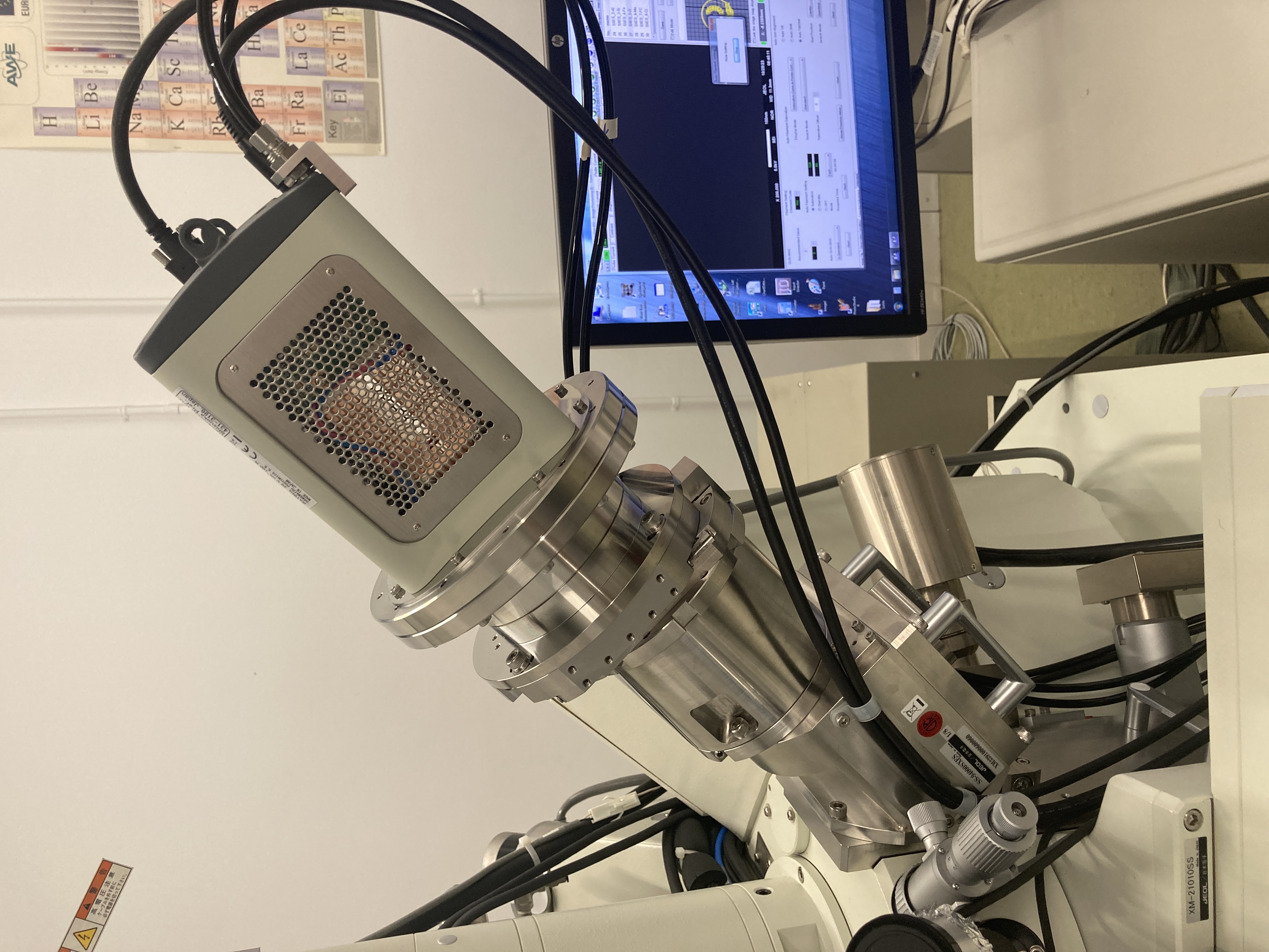
ARPES Angle Resolved Photoelectron Spectroscopy
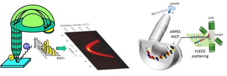
ARPES allows to measure directly the electronic band structure of crystalline solids. Electrons are detected retrieving information about initial state energy, momentum and spin.
UPS Ultra Violet Spectroscopy
Ultraviolet photoelectron spectroscopy (UPS), also described as photoemission spectroscopy (PES) if applied to measurements on solid surfaces, is suitable for measuring spectral features close to the Fermi level for surfaces or adsorbates, and particularly useful for determining work function and electronic properties of valence band of a material.
SXPSM Scanning XPS Microprobe
ESFOSCAN equipment performs XPS with monochromatic, micro-focused, scanning X-ray source. The X-ray induced secondary electron imaging provides a perfect correlation between imaged areas and spectroscopy. Moreover, UPS and LEIPS measurements can be performed at the same location to characterize the electronic band structure (VB and CB parameters).

