Due to the upcoming SLS 2.0 and ELETTRA 2.0 upgrade, this technique will be unavailable at PSI, ELETTRA and CNR-IOM(TS) until further notice.
XAS is a technique for determining the local electronic, structural and magnetic properties of matter in different scientific fields including molecular and condensed matter physics, material science, engineering, chemistry, earth science and biology.
When the incident photon energy is scanned through the energy of a core shell, an abrupt increase in the absorption cross-section, the so-called absorption edge representing a particular core shell, makes XAS an element and orbital selective technique.
XAS is very sensitive to the local bonding environment of the absorbing atom, providing information on oxidation state or density of states and/or local orbital symmetry.
XAS measurements are polarization dependent. Linearly polarized light can be used to determine the orbital occupancy or to look for the direction of chemical bonds of the atom selected by its absorption edge. This is particularly suited for covalent bonds like in molecules or polymers.
The experiment is performed at synchrotron radiation sources, which provide intense and tunable X-ray beams.
The XAS spectra can be measured in fluorescence or in electron yield modes.
Fluorescence yield spectra can be acquired in transmission measuring the incoming and transmitted light through a sample, or in reflection detecting the photons that are emitted after the electrons of lower binding energy fill the created core hole.
Electron Yield spectra are collected by measuring the photoelectrons created by the absorbed X-rays through excitations of core electrons to empty states above the Fermi level. The created holes are filled by Auger decay. The intensity of the emitted primary Auger electrons is a direct measure of the X-ray absorption process (Auger Electron Yield) which is strongly surface sensitive. As they leave the sample, the primary Auger electrons create scattered secondary electrons which dominate the total electron yield (TEY) intensity.
Elettra synchrotron, Apple II undulators; variable polarization (horizontal, vertical, circular ±); beam size on the sample 350x350 (HxV, µm2), vertical size can be reduced on request, flux on sample @10 µm slits (best resolution) (ph./s) 2x10¹²-6x10¹⁰
Hamamatsu fast fluorescence detector; photodiode, setup for x-ray absorption transmission measurement, drain current for total electron yield, Scienta R3000 analyzer for partial electron and Auger yield; setup for measurements in static liquid environment
35-1650 eV
(E/dE) 20000-5000
Many samples can be accommodated in a 25x25 mm2 area; T range: 50-1200 K (PID-controlled)
Base pressure: UHV
Heating stages (ebeam, direct current, PBN), ion gun (VG), 4 evaporator ports (CF40), gas inlet valve (variable leak valve), diamond file scraper, cleaver, LEED (OciLEED); evaporators for organic molecules; e-beam evaporator (Omicron) for metals (evaporation at low sample temperatures is also possible)
ARPES and XPS are possible in the same chamber; 4 degree-of-freedom manipulator;. x-ray magnetic circular dichroism (max 0.5 T, measurements in remanence); possibility to apply electric fields (measurements in remanence); possibility to superimpose synchrotron beam to laser beam for pump-probe measurements (100 ps resolution)
no
na
0
CNR-IOM @TS
Italy
XAS - High Energy APE Beamline @ Elettra Synchrotron
na
XCMD, XLMD (X-ray linear magnetic dichroism), LMDAD (linear magnetic dichroism in the angular distribution)
Elettra synchrotron, Apple II undulator; variable polarization (horizontal, vertical, circular ±); beam size on sample 500x200 (HxV, µm2), entry slit reduction to 100x100 possible, zone plate optical condenser reduction to 1x3 possible
Omicron EA 125 analyser; drain current Keithley 6514 picoammeter (for XAS/XMCD/XMLD); 10x10 mm silicon photodiode; total yield 20mm channeltron
(E/dE) 5000
T range: 30K- 300K (He flow cryostat) for LT stage (while measuring), 300K-700K for HT stage (while measuring); magnetic field: up to 1000 Oe in pulse mode, up to 200 Oe in continuous mode
The microXAS beamline provides X-ray absorption spectroscopy (XAS) and X-ray fluorescence (XRF) experiments requiring high spatial resolution as well as investigations of time-dependent phenomena in the femtosecond time regime
Minigap in-vacuum undulator, polarization linear horizontal, flux on sample 2x10¹² ph/s/400mA, spot size on sample 1 x 1 µm2
Photon energy resolution: 0.02%
FEMTO endstation for ultrafast (femtosecond to picosecond) dynamics in laser-excited systems using x-rays as a primary tool
3- 23 keV
PSI
Switzerland
XRF, XAS (transmission, total electron yield) - PHOENIX Beamline @ Swiss Light Source Synchrotron
The PHOENIX (Photons for the Exploration of Nature by Imaging and XAFS) beamline produces a high flux of soft to hard X-rays from an elliptical undulator (APPLE II). Two different endstations at two branches provide the users the necessary tools to perform X-ray microspectroscopic measurements (µ-XAS and µ-XRF)
Elliptical undulator (APPLE II), flux at 3 keV 1 x 10¹¹ph/s/0.1%BW/0.4A , focused spot size on sample
2.5 µm x 2.5 µm
Thin Si-diode for higher energies or thin polyethylene foil, sputtered with Ni, for lower energies. The X-ray fluorescence is detected either by a single-element or a 4-element Si drift diode array
0.8- 8 keV
5 sample stages
PSI
Switzerland
XAS, XRF, XES - Super XAS Beamline @ Swiss Light Source Synchrotron
The SuperXAS beamline includes a large variety of detection systems and sample environments and is thus attractive for researchers from a variety of fields: e.g. material science, catalysis, environmental science, biology, geology and archeology. Techniques that are available at the beamline include time-resolved X-ray absorption fine structure (XAFS) spectroscopy (minute to the millisecond range), X-ray fluorescence (XRF) and X-ray emission spectroscopy (XES)
Super-bending, flux on sample: 1012 ph/s/400 mA, spot size on sample: 100x100μm2 to 5x0.5μm2
Mythen II, Pilatus 100 K
ΔΕ/Ε 2.0 x 10⁻⁴ for Si(111) and 0.5 x 10⁻⁴ for Si(311)
4.5 - 35 keV
SOLEIL
France
XAS, EXAFS - Samba Beamline @ Soleil Synchrotron
High and low temperature, reactor, wet cell
SOLEIL
France
Time resolved photoemission XPS, ARPES, XAS, XMCD - Tempo Beamline @ Soleil Synchrotron
Time resolution<50ps
SOLEIL
France
XPS, HR-ARPES, Spin-resolved PES - Cassiopee Beamline @ Soleil Synchrotron
Electronic structure determination using scanning-angle-resolved photoemission spectromicroscopy combined with local structural characterisation by means of photoelectron diffraction
Resolution <120nm
CNR-IOM @TS
Italy
XAS @ ALOISA beamline
na
Aloisa is a spectroscopic beamline (XPS, XAS, ResonantXPS) dedicated to Surface Science.
Systems: single crystals, Self Assembled Monolayers, small molecule adsorbates (poly- and hetero-aromatics), ultra-thin films.
Phenomena: on-surface synthesis and modification of molecular adsorbates, charge transfer at hybrid organic-inorganic interfaces, molecular orientation at surfaces.
Insertion Device:
- 1.5 m phase shifting Undulator (constant gap): 21 x 7cm periods.
- linear polarization.
- fully integrated in the acquistion software suite.
Monochromator:
- type SX-700 (plane mirror-grating) with collimated beam (2x Paraboloidal Mirrors at 0.5˚ grazing incidence).
- multiple working curves for optimization of flux, resolution, rejection of higher orders (depending on specific needs).
- linear polarization.
- fully integrated in the acquistion software suite.
- VERY HIGH TRANSMISSION AT CARBON IONIZATION THRESHOLD (40-50%).
- Flux at Sample: ~1x10^12 ph/sec from 200 to 1000 eV in working condition (Exit Slits = 20µm).
- photon energy resolving power (E/DE): >5000 from 200 to 900 eV in working condition (10^12 ph/sec).
XAS: partial electron yield by means of a channeltron equipped with a polarizable grid for low energy secondary electrons rejection (NO drain current measurement available). Simultaneous measurement of drain current on refocussing mirror (i0) for flux normalization and absolute energy calibration at the C, N, O K-edge and Fe, Cr L-edge.
XPS: homemade hemisperical analyzer (mean radius 66mm); 2xMCP + 2D-DelayLine detector for very high dynamical range (1 MHz);
PED: fully rotatable analyzer with small detection angle (<2˚ FWHM);
ResPES: undulator, monochromator, manipulator and analyzer movements fully integrated in the acquistion software suite.
FULLY AUTOMATED ACQUISTION SOFTWARE (Labview suite).
XPS: photoelectron in the 50-1500 eV range
XPS:
- electron energy resolution: 1% Pass Energy (minimum 35 meV)
- standard working conditions: 10-30 eV Pass Energy
Solid samples with polished surface (reflective) for sample alignment (phosphorum plat with TV camera at the end of experimental station for checking the reflected beam.
Sample holders (POD) with on-board heating system (radiative or electron bombardment), LN2 cooling, two thermocouples.
Sample size: max thickness 3 mm; max width 12-14 mm; min width 4 mm.
Minimum temperature: 150 K
Max temperature: 1050 K (flash).
PID temperature control with programmable ramps.
Real time XPS during heating from 150 to 650 K.
UHV experimental chamber (10^-11 mbar); UHV preparation chamber (10^-10 mbar).
Praparation chamber in-line between exp. chamber and refocussing mirror (UHV must be recovered before measurements.
Manipulator travelling forth and back between exp. and prep. chambers.
Max partial pressure for gas dosing 10^-6 mbar.
MBE cryovessel (H2O) with four slots for evaporators, shutters and microbalances.
Homemade Knudsen cells (two crucibles, either BN or quartz) with inner thermocouple for organic molecules and low vapour tension matyerials (up to 600˚C).
Omicron e-beam cells for metal evaporation.
Standard UHV equipment for sample cleaning and ordering.
Dedicated software (freeware) for data analysis and fittings (XPS, NEXAFS, ResPES 2D maps) based on IgorPro platform.
no
na
0
Monochromator energy range: 130-1500 eV.
Undulator First Harmonic:130-450 eV at B.E. = 2.0GeV (140-550 eV at B.E. = 2.4GeV)
MANIPULATOR:
- fully motorized six-degrees of freedom with high resolution (<0.01˚ on three rotations; <0.01mm on three translations).
- horizontally mounted (coaxial to the photon beam) for grazing incidence measurements.
- rotations and translations fully integrated in the acquisition software suite for surface scanning (to prevent radiation damage).
XAS: change of surface orientation with respect to polarization from Transverse Magnetic (p-pol) to Transverse Electric (s-pol) by sample rotation around the photon beam at constant grazing angle.
XPS: standard XPS at grazing incidence (4˚) and normal emission for high signal yield;
fully rotatable analyzer (zenithal angle: 0˚-100˚) for any surface azimuth (±95˚) and surface orientation w.r.t. polarization;
analyzer rotation fully integrated in the acquistion software suite.
Photon beam transverse size at sample position (h x w): 30-50 µm x 150 µm.
RHEED.
Microbalances integrated in the MBE system.
Not allowed to Users
KIT
Germany
XAS at KIT: Soft X-Ray Spectroscopy, Microscopy, and Spectromicroscopy (WERA)
The soft x-ray analytics facility WERA provides a coherent combination of electron spectroscopies and microscopies for studying in detail the chemical (electronic) and magnetic structure of bulk materials, thin films, and micro- and nanostructured objects.
Methods for Electron spectroscopy and spectromicroscopy: XAS, PES (XPS), SXMCD, µ-XAS, µ-PES (±-XAS), µ-SXMCD, topography
Equipment: Three experimental stations at WERA equipped with PEEM, electron energy analyzer, detectors, cryostats, sample preparation chambers, loadlocks, in-vacuo sample transfer etc.
AU-ISA
Denmark
NEXAFS- MatLine beamline @ASTRID2
na
MatLine is dedicated to the electronic properties of surfaces and interfaces on metal, metal oxide and semiconductor, especially of adsorbed molecules on substrates.
Multi-pole wiggler from ASTRID2; Resolving power 200 to 3,500; Typical Flux 1x10¹¹ photons/sec; Spot size: 0.7 mm X 0.7 mm.
SPECS PHOIBOS 150 1D-DLD electron analyzer
Temperatures in the range of 110K – 1200K; Non-insulating samples at size less than 12mm X 7mm X 2mm; Possibility of measuring certain type of powders
Measurements are carried out under UHV
2 evaporator ports (CF40), installation without break main vacuum; gas inlet; load-lock; auto heating while measuring; LEED; ion gun; RESPED is not available at the moment
no
na
0
20-700 eV, with the possibility up to 1260eV at lower resolution and flux
XPS is a surface spectroscopic technique for quantitative measurements of the elemental composition or stoichiometry and the chemical state of the present elements, like their oxidation state and chemical bonds. XPS is highly surface sensitive, giving chemical and binding energy information from the a narrow region close to the surface.
XRD provides non-destructive information on the structural order of a material. At large scattering angles XRD permits to identify different crystal phases and to quantify lattice distances and crystalline volume fractions. At low angles of incidence the surface roughness of a single crystal and the thickness of a deposition layer can be obtained.
In SEM a beam is scanned over a sample surface while a signal from secondary or back-scattered electrons is recorded. SEM is used to image an area of the sample with nanometric resolution, and also to measure its composition, crystallographic phase distribution and local texture.
In TEM/Scanning TEM (STEM) high energy electrons incident on ultra-thin samples, allow imaging, diffraction, electron energy loss spectroscopy and chemical analysis of solid materials with a spatial resolution on the order of 1-2 Å. Samples must have a thickness of a few tens of nanometres and are prepared in sample preparation laboratory.
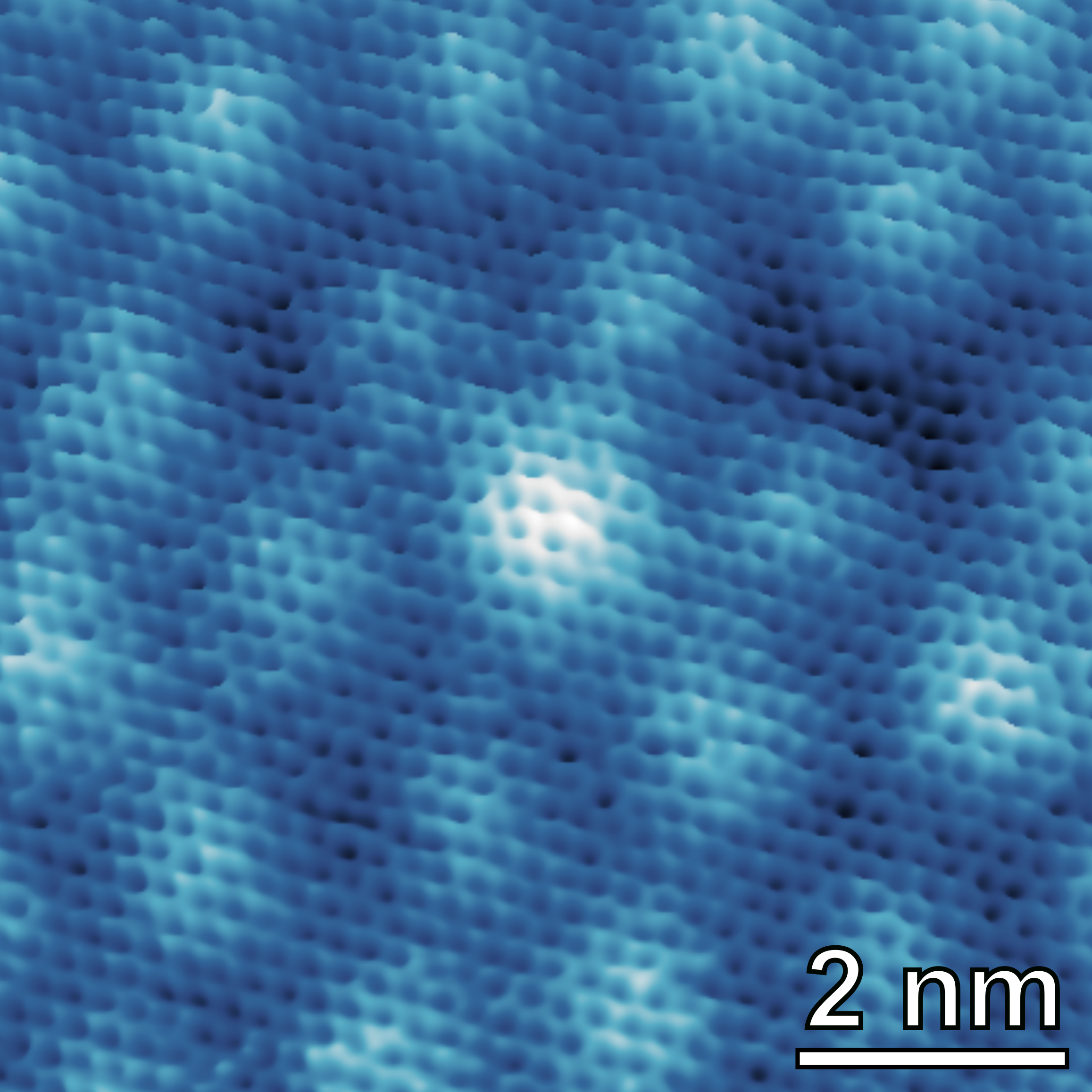
STM allows imaging conductive surfaces at the atomic scale. It is possible to characterize the distribution of surface terraces and steps, as well as to determine the atomic arrangement of (ordered) surface (over)structures.