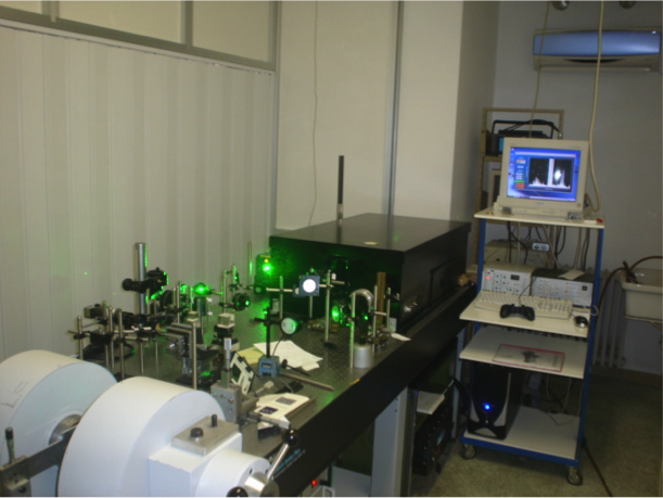
Three Brillouin Light Scattering (BLS) experimental setups are available:
1) Conventional Brillouin light scattering (BLS) setup, which allows BLS measurements from thermally excited spin waves in the frequency range between 1 and 200 GHz and a spatial resolution of 30 um. Possibility to apply static magnetic field up to 2 T parallel to the sample surface and to change the incidence angle of light with respect to the sample normal. This allows to explore a wave vector range from 0 to2.2x10^7 rad/m and to measure the dispersion of spin waves (frequency vs wave-vector). Azimuthal rotation of the sample around its normal is also possible to investigate the presence of in-plane magnetic anisotropy.
2) Micro-focused Brillouin light scattering with a lateral resolution down to about 250 nm intensity of the applied magnetic field up to 1 T (out-of-plane) and 0.25 t in-plane. This technique allows to perform 1D and 2D spatial maps of the spin-wave intensity generated by either dc current or a microwave current (up to 20 GHz).
3) Combined micro-Brillouin and micro-Raman set-up to observe collective dynamics (mechanical characterization) by BLS and high frequency molecular vibrational modes (chemical characterization) by Raman in a variety of samples with u-metric spatial resolution (Lateral spatial resolution 500 nm up to 2 um). Frequency resolution: in Brillouin spectra 100 MHz, in Raman spectra 2 cm^(-1)). Laser source at 532 nm. The temperature range covers from -195°C to 300°C. Plus supporting equipment, such as an Atomic Force Microscope operating both in contact and tapping mode and capable of Magnetic force microscopy measurements, and a magneto-optic kerr effect magnetometry apparatus with photo-elastic modulator operating at 50 kHz and lock-in amplification, for measuring hysteresis loops of magnetic nanostructures in the longitudinal configuration and maximum applied field of 200 mT.
Brillouin Light scattering spectroscopy from spin waves
Study of the spin waves excitation in low dimensional ferromagnetic nanostructures
Single mode laser operating at 532 nm
Frequency range: 0-200 GHz
Magnetic field range up to 2 T (on 3mm poles gap)
Wavevector range: 0- 2.2 10^7 m^-1
0.1 GHz
30-40 microns
3D translational stage
Incidence angle of light (θ) from 0 to 90 degrees (Explored wave vector range from 0 to 2.2x10^7 rad/m)
Azimuthal angle (φ) from 0 to 360 degrees
Maximum dimensions 1x1 cm^2
Room temperature
Reversable magnetic field applied in the sample plane up to 1.5 T for Magnetostatic surface configuration (Damon-Eshbach geometry) and 0.2 T in the backward configuration.
Optical microscope to find patterned areas prepared on the same substrate
CNR-IOM (PG)
Italy
Micro-BLS
200 mW, single-frequency, Excelsior Diode-Pumped Solid-State (DPSS) laser, operating in the spectral line of 532 nm, with a line width of approximately 10 MHz.
Power on the sample about 10 mW
0.1GHz
250 nm
A nanoposition stage allows to position the sample with a precision down to 10 nm on all three axes.
Room temperature
In-plane magnetic field of 0.25 T
Field projected out-of-plane of 1T
A coaxial viewing system based on a collimated LED light source (455 nm wavelength), a beam expander, and a CCD camera is used to obtain a direct visualization of the laser spot and of the
sample region under investigation.
6220 Keithley precision current source
2182A Leithley nanovoltmeter
Agilent EXA N9010A Signal Analyzer 10 Hz-26.5 GHz
Agilent E8257D Analog Signal Generator 250 kHz-20 GHZ
CNR-IOM (PG)
Italy
Combined micro-Brillouin and micro-Raman set-up
Mechanical characterization at the microscale
laser at 532 nm P=100mW
-80 to 80 GHz
100 MHz
1 um
(x,y,z) inverted microscope range (1cm,1 cm,1 cm) with a step of 1 um
A coaxial viewing system based on a white LED light source, a beam expander, and a CCD camera is used to obtain a direct visualization of the laser spot and of the sample region under investigation.
AFM is a surface sensitive technique permitting to obtain a microscopic image of the topography of a material surface and certain properties (like friction force, magnetization properties…). Typical lateral image sizes are within a range of only a few Nanometers to several Micrometers, and height changes of less than a Nanometer.
In TEM/Scanning TEM (STEM) high energy electrons incident on ultra-thin samples, allow imaging, diffraction, electron energy loss spectroscopy and chemical analysis of solid materials with a spatial resolution on the order of 1-2 Å. Samples must have a thickness of a few tens of nanometres and are prepared in sample preparation laboratory.
In SEM a beam is scanned over a sample surface while a signal from secondary or back-scattered electrons is recorded. SEM is used to image an area of the sample with nanometric resolution, and also to measure its composition, crystallographic phase distribution and local texture.
XRD provides non-destructive information on the structural order of a material. At large scattering angles XRD permits to identify different crystal phases and to quantify lattice distances and crystalline volume fractions. At low angles of incidence the surface roughness of a single crystal and the thickness of a deposition layer can be obtained.
When X-ray absorption is measured with circularly/linearly polarized x-rays, spin and angular momenta can be determined in ferromagnetic/antiferromagnetic systems, respectively. Dichroic effects arise by the difference between spectra measured with different helicity/polarization orientation of the X-ray photons.