X-ray Magnetic Circular/Linear Dichroism
Electronic & Chemical & Magnetic Characterization (X-ray/soft-X-ray spectroscopy)
Due to the upcoming SLS 2.0 and ELETTRA 2.0 upgrade, this technique will be unavailable at PSI and CNR-IOM(TS) until further notice.

@
provided at Large Scale Facilities by:
Instruments datasheets
CNR-IOM @TS
Italy
XAS, XMCD - BACH Beamline @ Elettra Synchrotron
x-ray spectroscopy with selectable polarization, x-ray magnetic linear/circular dichroism, time-resolved phenomena, out-of-equilibrium phenomena (pump-probe)
Elettra synchrotron, Apple II undulators; variable polarization (horizontal, vertical, circular ±); beam size on the sample 350X350 (HxV, µm2); Vertical size can be reduced on request, flux on sample @ 1 µm slits (best resolution) (ph./s) 2x10¹²-6x10¹⁰
Hamamatsu fast fluorescence detector; photodiode, setup for x-ray absorption transmission measurement, drain current for total electron yield, Scienta R3000 analyzer for partial electron and Auger yield; setup for measurements in static liquid environment
Heating stages (ebeam, direct current, PBN), ion gun (VG), 4 evaporator ports (CF40), gas inlet valve (variable leak valve), diamond file scraper, cleaver, LEED (OciLEED); evaporators for organic molecules; e-beam evaporator (Omicron) for metals (evaporation at low sample temperatures is also possible)
ARPES and XPS are possible in the same chamber; 4 degree-of-freedom manipulator. x-ray magnetic circular dichroism (max 0.5 T, measurements in remanence); possibility to apply electric fields (measurements in remanence); possibility to superimpose synchrotron beam to laser beam for pump-probe measurements (100 ps resolution)
CNR-IOM @TS
Italy
XMCD - High Energy APE Beamline @ Elettra Synchrotron
XCMD, XLMD (X-ray linear magnetic dichroism), LMDAD (linear magnetic dichroism in the angular distribution)
Elettra synchrotron, Apple II undulator; variable polarization (horizontal, vertical, circular ±); beam size on sample 500x200 (HxV, µm2), entry slit reduction to 100x100 possible, zone plate optical condenser reduction to 1x3 possible
Omicron EA 125 analyser; drain current Keithley 6514 picoammeter (for XAS/XMCD/XMLD); 10x10 mm2 silicon photodiode; total yield 20mm channeltron
T range: 30K- 300K (He flow cryostat) for LT stage (while measuring), 300K-700K for HT stage (while measuring); magnetic field: up to 1000 Oe in pulse mode, up to 200 Oe in continuous mode
PSI
Switzerland
XMCD - X-Treme Beamline @ Swiss Light Source Synchrotron
The X-Treme beamline is dedicated to x-ray magnetic (circular or linear) dichroism technique in the soft x-ray range. The technique is element selective and is used for example for the study of magnetic anisotropy and exchange coupling. The energy range covers the L2,3-edges (2p to 3d transition) of 3d transition metals and M4,5-edges of lanthanides (3d to 4f transition), in addition to the K-edges of light elements like O, N, F. Scientific areas of interest are: single molecule magnets, magnetic nanocrystals, self-assembly of nanomagnets on surfaces and strongly correlated electron systems
Elliptical polarizing undulator, linear (0 to 90o) or circular (left/right), flux (700eV): 4x10¹⁵ photons/s/0.1%BW/0.4 A, focused spot size: exit slit µm x 230 µm (V x H)
SOLEIL
France
Time resolved photoemission XPS, ARPES, XAS, XMCD - Tempo Beamline @ Soleil Synchrotron
KIT
Germany
XMCD/XMLD at KIT: Soft X-Ray Spectroscopy, Microscopy, and Spectromicroscopy (WERA)
The soft x-ray analytics facility WERA provides a coherent combination of electron spectroscopies and microscopies for studying in detail the chemical (electronic) and magnetic structure of bulk materials, thin films, and micro- and nanostructured objects.
Methods for Electron spectroscopy and spectromicroscopy: XAS, PES (XPS), SXMCD, µ-XAS, µ-PES (±-XAS), µ-SXMCD, topography
Equipment: Three experimental stations at WERA equipped with PEEM, electron energy analyzer, detectors, cryostats, sample preparation chambers, loadlocks, in-vacuo sample transfer etc.
@
provided at Large Scale Facilities by:
Also consider
Structural & Morphology Characterization
STM Scanning Tunneling Microscopy
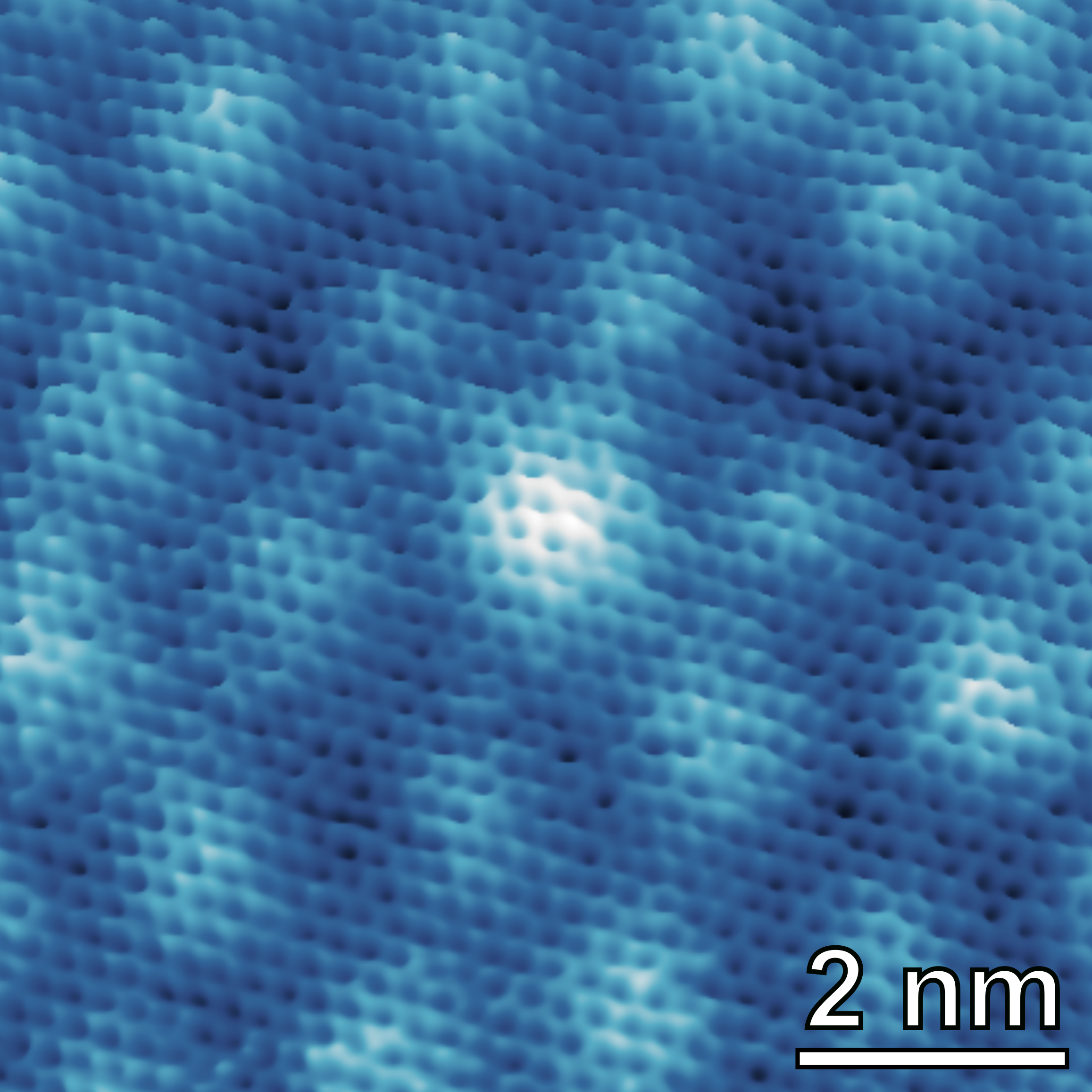
STM allows imaging conductive surfaces at the atomic scale. It is possible to characterize the distribution of surface terraces and steps, as well as to determine the atomic arrangement of (ordered) surface (over)structures.
Theory & Simulation
SGSEP Structural and ground-state electronic properties

This technique offers the possibility of simulating structural and electronic properties based on the electronic ground state, including electronic charge analysis, energetics of formation, structural and vibrational properties; IR, Raman, EPR, NMR, core-level XAS & XPS, STM & AFM.
Electronic & Chemical & Magnetic Characterization
XAS X-ray Absorption Spectroscopy

XAS is sensitive to the local bonding environment of the atom absorbing the X-rays, providing information on oxidation states, local orbital symmetry, molecular orientation and chemically selective density of states. It is widely used in molecular and condensed matter physics, material science, engineering, chemistry, earth science and biology.
Electronic & Chemical & Magnetic Characterization
XPS X-ray Photoelectron Spectroscopy

XPS is a surface spectroscopic technique for quantitative measurements of the elemental composition or stoichiometry and the chemical state of the present elements, like their oxidation state and chemical bonds. XPS is highly surface sensitive, giving chemical and binding energy information from the a narrow region close to the surface.
Structural & Morphology Characterization
XRD X-Ray Diffraction

XRD provides non-destructive information on the structural order of a material. At large scattering angles XRD permits to identify different crystal phases and to quantify lattice distances and crystalline volume fractions. At low angles of incidence the surface roughness of a single crystal and the thickness of a deposition layer can be obtained.

