Fluorescence Spectroscopy

Electronic & Chemical & Magnetic Characterization (Luminescence spectroscopy)

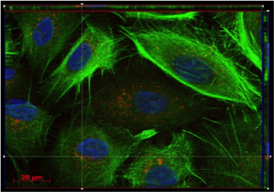
Fluorescence is a type of photoluminescence where light raises an electron to an excited state. The excited state undergoes rapid thermal energy loss to the environment through vibrations, and then a photon is emitted from the lowest-lying singlet excited state. This process of photon emission competes for other non-radiative processes including energy transfer and heat loss. Fluorescence spectroscopy uses a beam of light that excites the electrons in molecules of certain compounds, and causes them to emit light. That light is directed towards a filter and onto a detector for measurement and identification of the molecule or changes in the molecule.

The basic function of a fluorescence spectrometer is to irradiate the specimen with a desired and specific band of wavelengths, and then to separate the much weaker emitted fluorescence from the excitation light. In a configured fluorescence microscope in particular, only the emission light will reach the eye or detector so that the resulting fluorescent structures are superimposed with high contrast against a very dark background. Through the use of multiple fluorescence labelling with different probes can identify several target molecules simultaneously. 

The Fluorescence process, allows users through microscopy to determine the distribution of a single molecule species, its amount and its localization inside materials, devices, cells and co-localization with other molecules. When the reflected light and background fluorescence is filtered in this type of spectroscopy/microscopy the targeted parts of a given sample can be imaged.

i
@
          provided at NFFA-Europe laboratories by:
@
          provided by:
EURONANOLAB
France

Instruments datasheets

CIC biomaGUNE
Spain
Edinburgh FS5
JRC - ISPRA
Italy
Fluorescence Microscopy
Fluorescence (confocla) microscopy of live cells uses either fluorescent proteins or cell membrane-permeable, non-toxic fluorescent stains to monitor biological processes involved upon ENMs exposure.
EURONANOLAB
France
FS at EURONANOLAB - NANOTEC
EURONANOLAB
France
FS at EURONANOLAB - PoliFAB
i
@
          provided at NFFA-Europe laboratories by:
@
          provided by:
EURONANOLAB
France

Also consider

Structural & Morphology Characterization

AFM Atomic Force Microscopy

AFM is a surface sensitive technique permitting to obtain a microscopic image of the topography of a material surface and certain properties (like friction force, magnetization properties…). Typical lateral image sizes are within a range of only a few Nanometers to several Micrometers, and height changes of less than a Nanometer.

Structural & Morphology Characterization

LSCM laser scanning confocal microscopy

This technique has the mission of enabling researchers to visualise and to monitor cellular events in real time and in vivo down to the molecular level, enabling prolonged observations that are not possible with classic confocal microscopes and allowing unparalleled detail in soft matter imaging.

Structural & Morphology Characterization

SEM Scanning Electron Microscopy

In SEM a beam is scanned over a sample surface while a signal from secondary or back-scattered electrons is recorded. SEM is used to image an area of the sample with nanometric resolution, and also to measure its composition, crystallographic phase distribution and local texture.

Electronic & Chemical & Magnetic Characterization

XPS X-ray Photoelectron Spectroscopy

XPS is a surface spectroscopic technique for quantitative measurements of the elemental composition or stoichiometry and the chemical state of the present elements, like their oxidation state and chemical bonds. XPS is highly surface sensitive, giving chemical and binding energy information from the a narrow region close to the surface.

Structural & Morphology Characterization

TEM Transmission Electron Microscopy

In TEM/Scanning TEM (STEM) high energy electrons incident on ultra-thin samples, allow imaging, diffraction, electron energy loss spectroscopy and chemical analysis of solid materials with a spatial resolution on the order of 1-2 Å. Samples must have a thickness of a few tens of nanometres and are prepared in sample preparation laboratory.