Though based on the same physical phenomena regarding the interaction between radiation and matter, today’s IR spectrometers have evolved in the way they irradiate the sample, replacing the former monochromators for interferometers, giving rise to the faster FT-IR spectroscopy.
Besides its classical application in chemical characterization, the use of infrared radiation has evolved giving rise to new techniques that go further:
VCD (Vibrational Circular Dichroism) is a technique that gives 3D information of molecules. It can be applied for determining the secondary structure of proteins and peptides, the purity of enantiomers and also their absolute configuration by comparison with previously reported data or with data obtained through theoretical simulations.
PM-IRRAS (Polarization Modulation-IR Reflection-Adsorption Spectroscopy) is a very useful technique for the analysis of ultrathin layers and coatings, monolayers and submonolayers and biomolecules, deposited on surfaces, especially for conductors (Au, Cu, Pd, alloys, etc). It allows for the study of not only the composition but also the organization, conformation and orientation of molecules on a given substrate. In addition, it is also useful for analysing phenomena affecting such surfaces, as could be corrosion processes. Thanks to the characteristics of this technique, samples can be measured without reference, giving rise to spectra free from atmospheric interferences such as carbon dioxide and water vapour
FT-IR Microscopy which allows for visible inspection of samples and to obtain FT-IR spectra by coupling all-reflective Vis-IR microscopes to a FT-IR spectrometer. It is useful for performing chemical characterisation in concrete points, and also for obtaining chemical maps of larger areas, with minimum spot sizes of 25-30 μm using conventional IR sources. Diffraction-limited resolution can be achieved exploiting the brightness advantage of IR Synchrotron Radiation (SR), for unveiling vibrational details at few microns in the Mid-IR regime. The non-damaging nature of IR SR and the use of IR-transparent fluidic devices permit the analysis of hydrated species, to follow sample dynamics and to work under physiological conditions at single cell level.
FT-IR Imaging is a very versatile toll for chemical imaging of large sample areas, that takes advantage from bi-dimensional Focal Plane Arrays (FPA) detectors. The technique is ideally suited for the rapid analysis of a large variety of chemically heterogeneous samples, from slice tissues to polymer blends, in transmission, reflection and ATR mode.
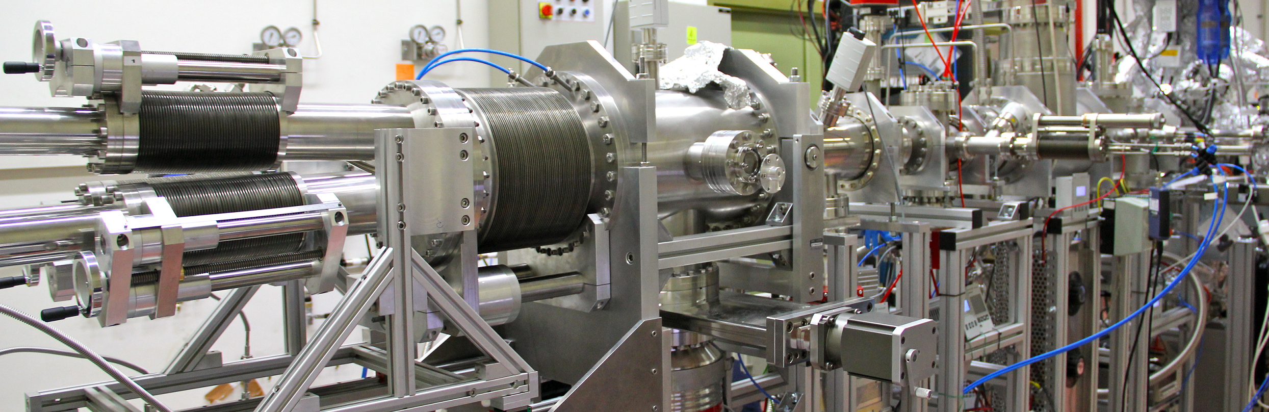
VERTEX 70v interferometer and Hyperion 3000 Vis/IR microscope @ Chemical and Life-Sciences branches of SISSI beamline at Elettra
na
Microscopy and Imaging in the MIR regime and with the newest VERTEX FM technology, for spectroscopy covering the FIR/THz and MIR in a single scan.
a. Halogen for the Near Infrared (NIR)
b. Globar for the Mid Infrared (MIR)
a. KBr beamsplitter for MIR regime up to ~400 cm-1
b. CaF2 beamsplitter for NIR
c. Ultra-wide range MIR-FIR beamsplitter (6000-50 cm-1)
a: DTGS, MCT and wide range DLaTGS detectors for spectroscopy measurements in the MIR and MIR-FIR regimes
b: MCT-A, MCT-B and FPA detectors for microscopy and imaging in the MIR regime
a: Max spectral resolution: 0.5 cm-1
b: Max spatial resolution: diffraction limited (typically in the range of few microns in the MIR spectral range)
The branchline is well suited for biological and biomedical experiments, encompassing tissue imaging and single-cell analysis on both dehydrated and hydrated samples. Bulk materials and surface analysis can also be performed selecting the appropriate sampling techniques.
a: For spectroscopy
Transmission measurements on pellets and within Diamond Compression cells (available with 0.8 and 2 mm culet)
Demountable liquid-cell T-controlled for transmission measurements of liquids
Attenuated Total Reflectance (ATR) measurements
i. Single Reflection ATR with Germanium, diamond and silicon IREs
ii. Grazing angle single reflection ATR with Ge IRE
iii. Multiple ATR with 25 reflection trapezoid IREs (Ge, Si, ZnSe, ...)
4. T-controlled Single Reflection monolithic ATR accessory for MIR-FIR measurements
b: For Microscopy
Transmission measurements on thin samples (slices) (15X and 36X objectives)
Reflection/Transflection measurements
Grazing Angle Reflection measurements
Micro-ATR measurements
Room temperature and pressure microscopy measurements, with possibility to T-control from 10 to 90 °C. Spectroscopy measurements are possible also in vacuum (10-3 Torr) and in the same T-range.
a: Ready-to-use bio-compatible liquid cells for live cell analysis
b: Possibility to design ad-hoc chips for reflection and transmission measurements in collaboration with potential users
no
na
0
CSIC-ICMM
Spain
Stardust: INFRA-ICE module: Transmission
IR spectroscopy (mid- and near IR) in transmission under ultra-high vacuum conditions and at low temperatures (T=15-300K). Complete evacuated optical path to avoid background signals from gas phase species.
Bruker, VERTEX 70V spectrometer.
Mid-IR: standard globar and a water-cooled high-power source.
Near-IR: tungsten halogen lamp.
12800 cm^-1 - 850 cm^-1
For mid-IR: DLaTGS and MCT detectors.
For near-IR: InGaAs detector.
Maximum dimensions: 15 x 15 mm^2.
For low temperature experiments: 10 x 10 x 1 mm^2.
Substrate needs to be IR transparent (e.g., KBr, ZnSe, CsI...).
Base pressure: 4 x 10^-10 mbar. Temperature control: 15 - 300 K. Gases up to 10^-6 mbar can be introduced in the sample chamber.
Quadrupole mass spectrometry (1 - 200 amu) for Thermal Programmed Desorption experiments (T = 15 - 300 K).
No
12800 cm^-1 - 850 cm^-1
Maximum resolution: 0.16 cm^−1
UV irradiation (Ly-alpha line: 121.6 nm). Electron irradiation (1-500 meV). Ion irradiation (0.2 - 6 keV).
No
JRC - ISPRA
Italy
Micro-FTIR (Fourier Transform InfraRed Spectrometer coupled with a microscope)
FTIR stands for Fourier Transform InfraRed, a method of infrared spectroscopy. When IR radiation is passed through a material, some radiation is absorbed by the sample and some passes through (is transmitted). The resulting signal at the detector is a spectrum representing an absorption by molecular vibrations within the sample. The usefulness of infrared spectroscopy arises because different chemical structures (molecules, side groups, functionalities) produce different spectral fingerprints.
EURONANOLAB
France
FTIR at EURONANOLAB - MMI
na
na
no
na
0
no
EURONANOLAB
France
FTIR at EURONANOLAB - IMT
EURONANOLAB
France
FTIR at EURONANOLAB - CNR-Nanotec
EURONANOLAB
France
FTIR at EURONANOLAB - LAAS
TUG
Italy
FT-IR spectroscopy
FT-IR spectroscopy on liquids, pellets, foils, thin films
Bruker Alpha FTIR spectrometer
Spectral range: 7500 cm-1 a 350 1/cm; Maximal resolution: 21/cm; Signal/Noise ratio: > 55,000:1 (1 min measurement time, spectral resolution 4 1/cm)
polymers, coated surfaces, solutions.
ATR, transmission sample compartment with a 2x3" standard sample holder, external reflectance
ALBA
Spain
SR-FTIR Bruker Hyperion 3000
Modern synchrotron-based infrared spectrometer and microscope capability covering a wavelength range from about 1 µm to ∼100 µm with a spectral region optimized initially for investigation between 2.5-14 µm.
Transmission, Reflection, Attenuated total reflection (ATR) and Grazing incidence are the most important geometries for sample analysis, and are all available at the beamline.
We offer a multiple ion-cluster source operating in ultra-high vacuum devoted to the fabrication and characterization of highly controlled nanoparticles. They are produced in gas phase with high purity and controlled size, structure and stoichiometry and they can be collected in the desired coverage on arbitrary surfaces for different uses.
XPS is a surface spectroscopic technique for quantitative measurements of the elemental composition or stoichiometry and the chemical state of the present elements, like their oxidation state and chemical bonds. XPS is highly surface sensitive, giving chemical and binding energy information from the a narrow region close to the surface.
VUV/UV/Vis/NIR spectroscopy is the measurement of the attenuation of a beam of light after it passes through a sample or after reflection from a sample surface. It is useful to characterize absorption, transmission, and reflectivity of a variety of technologically important materials, such as gases, film, pigments, coatings, windows, and filters.
XRD provides non-destructive information on the structural order of a material. At large scattering angles XRD permits to identify different crystal phases and to quantify lattice distances and crystalline volume fractions. At low angles of incidence the surface roughness of a single crystal and the thickness of a deposition layer can be obtained.
SAXS is a non-destructive and versatile method to study the nanoscale structure of any type of material (solid, liquid, aerosols) ranging from new nanocomposites to biological macromolecules. Averaged particle sizes, shapes and distributions, porosity, degree of crystallinity and electron density maps with nanometer precision can be obtained.