Live cell imaging

Nano to Micro/Macro (in vitro assays and cell analysis)

The “Live Cell Imaging” facility is equipped with tunable fs laser excitation source modules (660nm-5000nm) and dual beam, dual raster-scanning modules, accompanied with 5 simultaneous detection channels, which are covering the excitation and detection of plethora of dyes or other fluorescent markers.

It is based on a fully motorized Zeiss Axio Observer microscope with an incubator (Temperature and CO2 control module) at the sample plane, which is appropriate for long observation times of living cells and tissues.

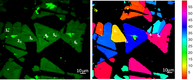
Apart from two-photon fluorescence (2pF) microscopy the custom-built Live Cell Imaging workstation is further equipped with polarization-resolved Second Harmonic Generation (P-SHG), polarization-resolved Third Harmonic Generation (P-THG) and Three-photon excited Fluorescence (3pF) imaging capabilities.

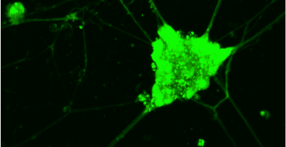
Consequently, except exogenous fluorescence markers (e.g. dyes) it can probe endogenous contrast agents which are appropriate e.g. for high resolution (<500nm) imaging of myofibroblasts, collagen type I, in fibrosis, wounds, joints, nanoliposomes etc.

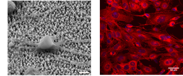
Additionally, the Live Cell Imaging facility provides experimental and theoretical tools for the quantitative imaging of scaffolds (used for example as implants) seeded with living cells or other biological material.

Finally, the Live Cell imaging workstation is further equipped with nanosurgery and fast calcium imaging capabilities

i
@
          provided at NFFA-Europe laboratories by:
FORTH
Greece

Instruments datasheets

FORTH
Greece
Live cell imaging
i
@
          provided at NFFA-Europe laboratories by:
FORTH
Greece

Also consider

Structural & Morphology Characterization

SEM Scanning Electron Microscopy

In SEM a beam is scanned over a sample surface while a signal from secondary or back-scattered electrons is recorded. SEM is used to image an area of the sample with nanometric resolution, and also to measure its composition, crystallographic phase distribution and local texture.

Nano to Micro/Macro

CCF Cell culture facilities

The Cell Culture Facility provides the necessary equipment (basic cell culture, sample preparation, functional assays and imaging equipment) for the study of the effect of biomaterials on specific cell behavior and function and cellular responses, such as cell survival, adhesion, morphology, proliferation, growth, migration and differentiation.

Nano to Micro/Macro

LSIVP Laser surface and in-volume Patterning

Laser patterning is a technique for the controlled patterning of materials at micro- and nano-scales. It offers the ability to directly write patterns on the surface and complex 3D channels into the bulk of solid materials, also biomaterials. Applications can range from microfluidic systems and sensors to tissue engineering scaffolds.

Structural & Morphology Characterization

NLM Nonlinear microscopy

NLM takes advantage of tightly focused ultra-short laser pulses (fs) to excite non-linear optical phenomena like Second Harmonic Generation, Third Harmonic Generation, Two-Photon and Three-photon excited Fluorescence. Along with laser raster-scanning of the sample, non-linear imaging microscopy of large areas is accomplished within seconds.

Structural & Morphology Characterization

CM Confocal microscopy

Confocal microscopy (CM) is an optical imaging technique that creates a virtual plane or slice, many micrometers deep within the analyzed sample. Compared to conventional microscopy, it provides fine detailed images of higher quality and with more contrast. In addition, virtual 3-D images of the analyzed microstructure can be obtained.