The “Nonlinear Microscopy” facility is based on a Zeiss Axio Observer Z1, fully motorized inverted microscope, which is coupled with tunable fs laser excitation source modules (660nm-5000nm) and dual beam, dual raster-scanning modules, accompanied with 5 simultaneous detection channels (in both forward and backwards detection geometries).
The polarization states of the excitation beams, as well of the polarization states of the detected nonlinear signals are fully controlled by combination of motorized retardation plates and polarizers.
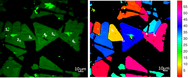
This allows the application of the state-of-the-art, advanced imaging techniques, namely Polarization-resolved Second Harmonic Generation and Polarization-resolved Third-harmonic (P-SHG and P-THG, respectively) imaging microscopies.
Along with theoretical tools that are based on nonlinear optics, the description and the interpretation of the produced nonlinear signals is offering insight at the nanoscale for the samples under examination.
The facility is further offering nonlinear microscopic imaging under cryogenic conditions (i.e. at 78°-300K°). This is accomplished by placing a cryostat in a special holder, at the sample plane of the nonlinear microscope.
@
provided at NFFA-Europe laboratories by:
@
provided at NFFA-Europe laboratories by:
Also consider
Structural & Morphology Characterization
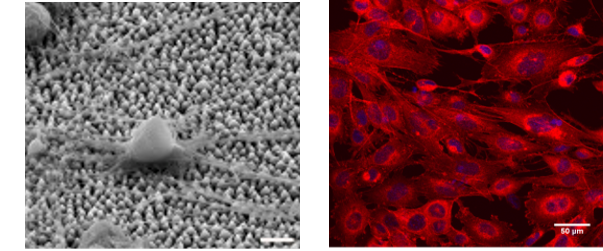
SEM Scanning Electron Microscopy
In SEM a beam is scanned over a sample surface while a signal from secondary or back-scattered electrons is recorded. SEM is used to image an area of the sample with nanometric resolution, and also to measure its composition, crystallographic phase distribution and local texture.
Nano to Micro/Macro
CCF Cell culture facilities
The Cell Culture Facility provides the necessary equipment (basic cell culture, sample preparation, functional assays and imaging equipment) for the study of the effect of biomaterials on specific cell behavior and function and cellular responses, such as cell survival, adhesion, morphology, proliferation, growth, migration and differentiation.
Nano to Micro/Macro
LCI Live cell imaging
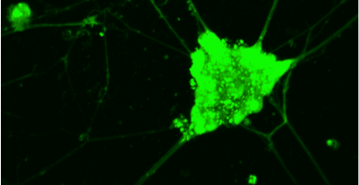
The “Live Cell Imaging” facility is equipped with advanced imaging microscopy techniques (based on two- or multi- photon excitation), which are appropriate for the all-optical minimally invasive, high-resolution (<500nm), deep (>500μm) monitoring of living cells and tissues for long periods of time.
Structural & Morphology Characterization
AFM Atomic Force Microscopy
AFM is a surface sensitive technique permitting to obtain a microscopic image of the topography of a material surface and certain properties (like friction force, magnetization properties…). Typical lateral image sizes are within a range of only a few Nanometers to several Micrometers, and height changes of less than a Nanometer.
Electronic & Chemical & Magnetic Characterization
RS Raman spectroscopy
Raman spectroscopy (RS) investigates the vibrational properties of a sample and provides chemical as well as structural information. RS does not require any specific sample preparation, size or condition and may be combined with micron/nano spatial resolution when operated using a confocal microscope/TERS or SNOM configuration.