Electron and ion beam technologies
IBA IBA (Ion Beam Analysis)
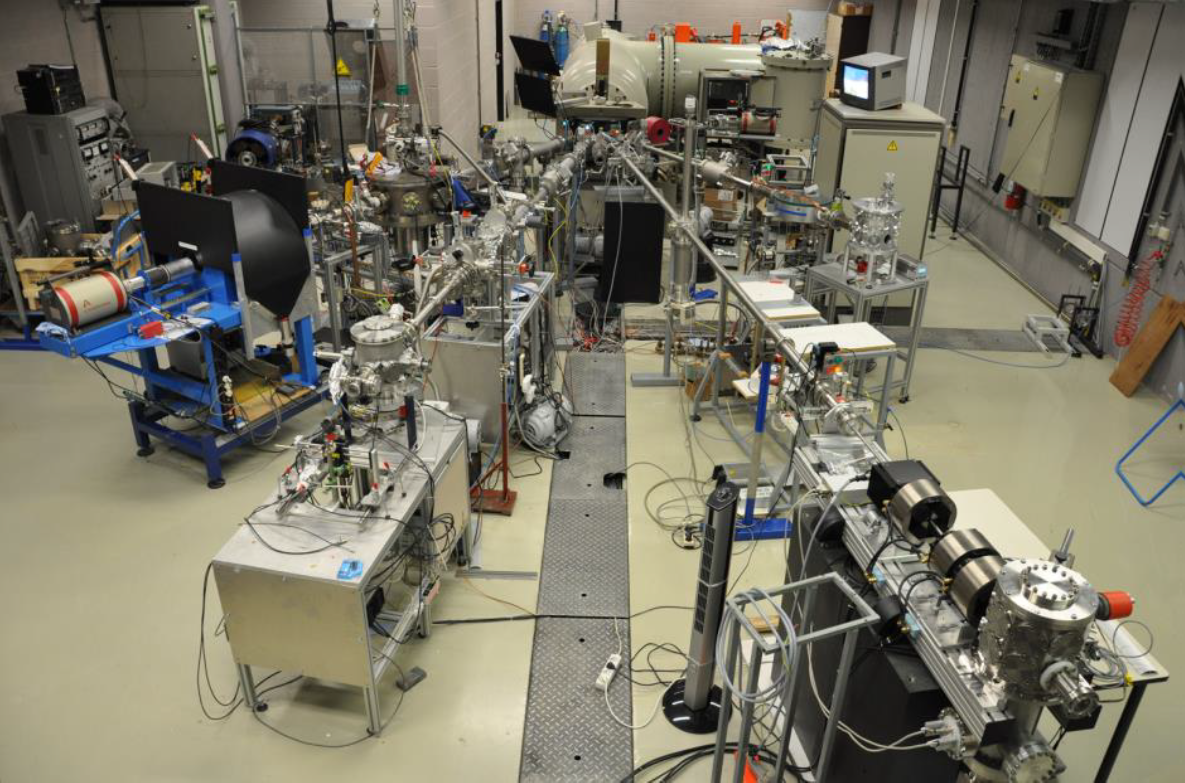
IBA consists of bombarding samples with few MeV ions to obtain information about depth profile and absolute element concentration (at/cm²) from a few nm up to a few µm. It combines different techniques that have their own features (light/heavy/trace element detection, etc.) but can be combined to work in a synergic way.
SIMS Secondary Ion Mass Spectrometry

By SIMS, samples are bombarded; sputtered and ionized atoms accelerate towards a mass spectrometer, providing surface mass spectra, images with lateral resolution in the 0.1 to 10 µm range and depth profiles with depth resolution in the 1 to 10 nm range. SIMS can be quantitative up to ppb sensitivity when reference samples are used.
FIB prep for TEM FIB sample preparation for TEM
In FIB an energetic beam of ions creates local removal of material by sputter erosion. Combination with an electron column, allows 2D and 3D nanometric structures to be achieved. Characterization by different detectors (SEM, BSE, STEM, X-Ray) is possible. FIB is the preferred technique to prepare extremely thin slices for TEM investigations.
APT Atom Probe Tomography

A voltage and/or a laser pulse is used to field evaporate atoms from the end of a specimen in the form of a sharp tip. By measuring the time-of-flight and the position of the evaporated atoms on a position sensitive detector a 3-D reconstruction of the position and chemical nature of detected atoms is possible with sub-nm resolution.
SEM Scanning Electron Microscopy

In SEM a beam is scanned over a sample surface while a signal from secondary or back-scattered electrons is recorded. SEM is used to image an area of the sample with nanometric resolution, and also to measure its composition, crystallographic phase distribution and local texture.
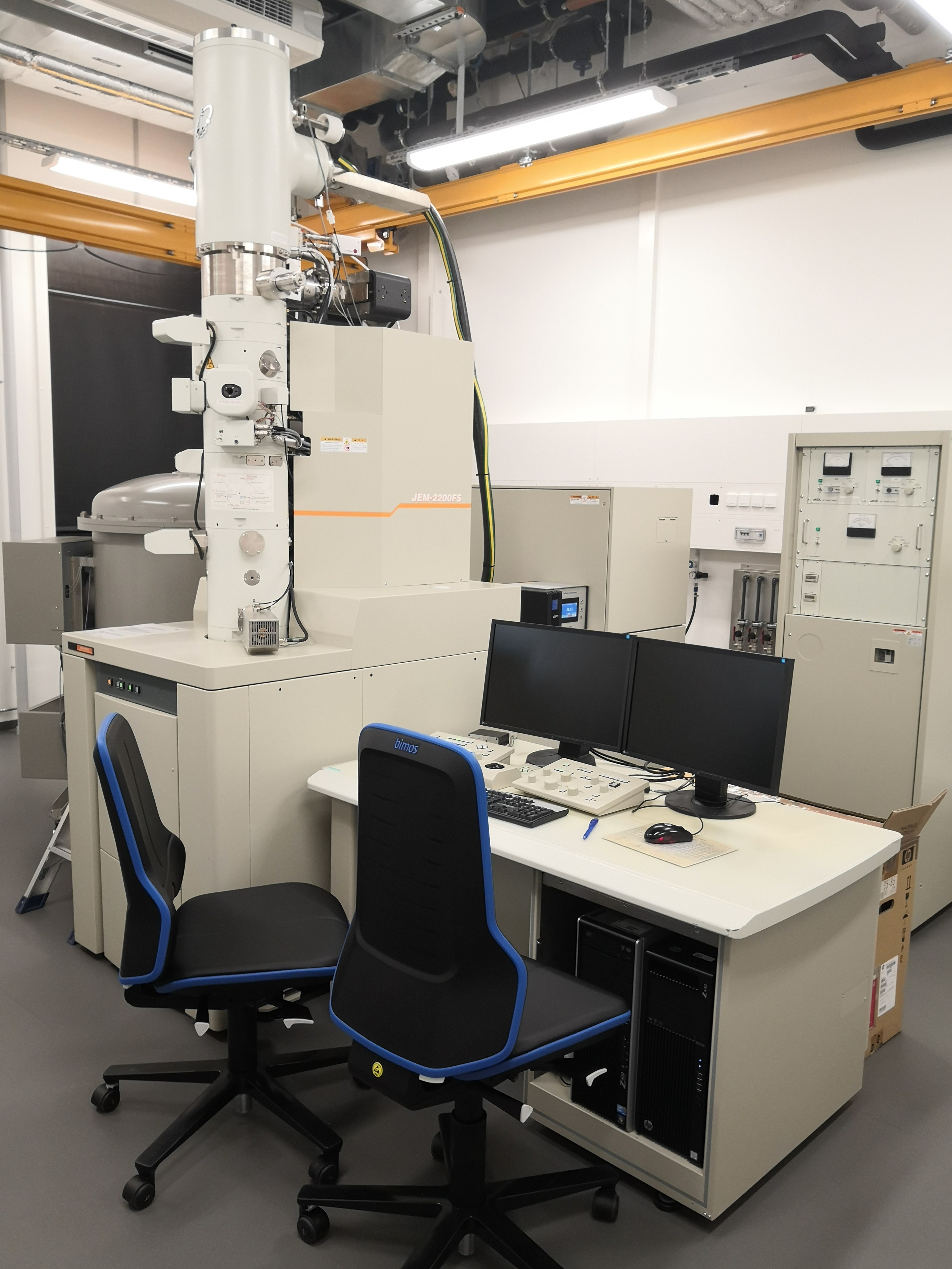
TEM Transmission Electron Microscopy

In TEM/Scanning TEM (STEM) high energy electrons incident on ultra-thin samples, allow imaging, diffraction, electron energy loss spectroscopy and chemical analysis of solid materials with a spatial resolution on the order of 1-2 Å. Samples must have a thickness of a few tens of nanometres and are prepared in sample preparation laboratory.
Cryo-TEM Cryo-Transmission Electron Microscopy
Cryo-Transmission electron microscopy is a technique for soft matter studies allowing real space investigations about shape and size distribution of particles, self-assembly and aggregation. Liquid sample can be directly observed on grid after blotting. An automatic plunge freezer is used to make cryo-specimen for direct imaging (Cryo-DI).
CLEM Correlative light-electron microscopy
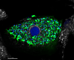
The Correlative Light and Electron Microscopy technique (CLEM) is a powerful approach that enables comprehensive investigations by combining optical microscopy, – using fluorochromes for biomolecule localization – and transmission electron microscopy (TEM), which provides high-resolution images at the nanoscale.

