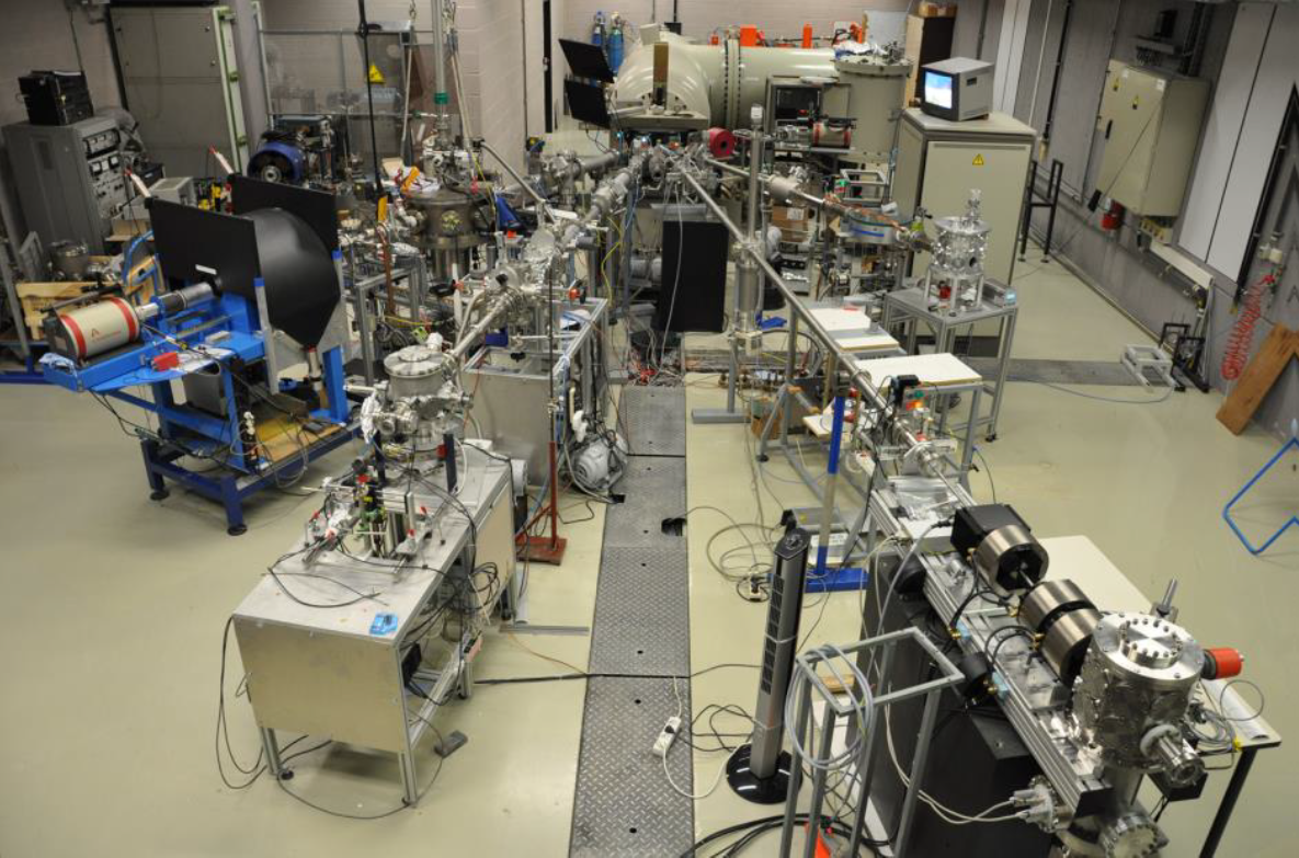
IBA (Ion Beam Analysis)
Structural & Morphology Characterization (Electron and ion beam technologies)
IBA consists of bombarding samples with few MeV ions to obtain information about depth profile and absolute element concentration (at/cm²). To achieve these properties, IBA combines different techniques, such as:
- RBS/EBS (Rutherford/Elastic Backscattered Spectroscopy): Consists in bombarding samples with light particles (protons or alphas) and detecting backscattered By the law of kinematics, only elements heavier sent particles could be detected. The main advantage of RBS/EBS is that it allows to determine the depth profile of the sample without needing to know exactly the sample composition (as it is the case for XRF).
Features:
-
- Elements: O-U
- Detection limit: 1 at. %
- Depth resolution: 10 nm
- Lateral resolution: 1 mm (broad beam)/ 1 µm (micro beam)
- ERDA (Elastic recoil Detection Analysis): Consists of bombarding sample with light particles (generally alphas particles) and detecting forward This technique allows notably to quantify (absolute value) Hydrogen in samples.
Features:
-
- Elements: H, B-Si
- Detection limit: 1 at. %
- Depth resolution: 15 nm
- Lateral resolution: 1,5 mm (broad beam)
- PIXE (Particles Induce X-ray Emission): Consists of bombarding sample with light particles (generally protons) and detecting (using a Germanium detector) X-rays emitted by element present in the sample. This technique is used to determine elements in samples and is very powerful to determine trace elements in a sample (1 Wt. ppm).
Features:
-
- Elements: Na-U
- Detection limit: 1 Wt. ppm
- Depth resolution: NA
- Lateral resolution: 1 mm (broad beam)/ 1 µm (micro beam)
- NRA (Nuclear Resonant Analysis): This technique uses nuclear reactions that happen at defined energy. These reactions present a high cross section, and due to energy loss of incident particle in the sample, it is possible to profile specific elements inside the sample. For example, the 1H(15N,ag)12C is very powerful in profiling hydrogen in samples with a depth resolution of around several nm and a detection limit of around tens of ppm of hydrogen. Note that in this reaction, gamma rays are generated and detected. The technique that uses nuclear reactions to generate and detect gamma is called PIGE (Particles Induce Gamma-ray Emission).
Features:
-
- Elements: H, B, C, N, F, O, …
- Detection limit: 001 at. %
- Depth resolution: 5 nm
- Lateral resolution: 1 mm (broad beam)/ 1 µm (micro beam)
All these techniques have their own particularities but can be realized at the same time (except for some NRA reactions) and work in a synergistic way (what we call Total-IBA). The size of the analyzing beam can vary from a few mm² to a few µm². One other advantage of IBA is that it is a non-destructive method. Generally, IBA is processed under vacuum condition (10-6 mbar) but PIXE can be made with extracted beam (meaning atmospheric pressure), that can be useful for samples that cannot be put under vacuum conditions.
Finally, in addition to these analysis techniques, our equipment allows to proceed to ion implantation (around any element in Mendeleev table, from a few keV until a few hundreds of keV) on an area around a few cm². The implanted dose, as well as the level of damage (amorphization) and implant profile, could also be controlled with our equipment.


