Correlative light-electron microscopy
Structural & Morphology Characterization (Electron and ion beam technologies)
The Correlative Light and Electron Microscopy technique (CLEM) is a powerful approach that enables comprehensive investigations by combining optical microscopy, – using fluorochromes for biomolecule localization – and transmission electron microscopy (TEM), which provides high-resolution images at the nanoscale. This allows for the correlation of data obtained from both methods, enhancing our comprehension of cellular processes. It enables the association of functional information with the specific structures in which these processes take place.
The Correlative Light and Electron Microscopy technique (CLEM) offers a comprehensive approach to studying biological specimens. It integrates fluorescence microscopy, using specific fluorochromes for biomolecule localization, with transmission electron microscopy (TEM), which provides high-resolution images at the nanoscale. This integration enables a deep understanding of cellular processes by associating functional information with ultrastructural data.
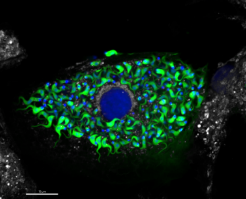
Fluorescence microscopy allows the simultaneous localization of multiple molecules, compartments, organelles, intracellular structures, as well as bacteria, viruses, and parasites, using specific fluorescent markers. Researchers can employ multiple fluorescent markers concurrently, typically between 4 to 5, facilitating informative, macro-level studies with a broader perspective. This versatility extends to various sample types, including live cells, fresh tissue specimens, and chemically fixed samples, all while preserving their fluorescence properties.
Achieving precise correlation of the same sample in Light and Electron Microscopy (CLEM) encompasses various methods which depend on the sample, the research goals and available technologies. Sample types can range from tissues, organs, cell cultures to cellular pellets, or subcellular fraction pellets, etc. Depending on the study, samples may be fresh, live, or chemically fixed. To ensure accurate localization between optical and electron microscopy, various localization techniques are utilized. These methods include growing cells on gridded coverslips with coordinates, fiducial-based localization, micropatterning, photo-oxidation, etc.
High-quality preparation methods used in electron microscopy allow the preservation of high-resolution structural information in cells. Depending on the goal of the study, CLEM can be applied to chemically fixed samples processed at room temperature, to cryo-sections of samples prepared according to Tokuyasu´s method or to samples processed by cryo-fixation and freeze substitution, being the latter the one that preserves the sample in a closest-to-native state. The subsequent physical sectioning of the sample in widths ranging from 50 to 300 nm produces much higher z-resolution data than can be obtained with confocal light microscopy of thick specimens and as the sections are thinner than the depth-of-field of the high-resolution objectives all structures can be imaged in focus. Therefore, the sample preparation is first observed in the fluorescent microscope which allows the detection of fluorescent particles smaller than the optical resolution. Afterwards, in the electron microscope, the same cell is located with the help of different landmarks (patterned grids, fiducial markers, sample profiles…), imaged (pictures or electron tomographic series that need to be afterwards computerised in a 3D electron tomogram) so the fluorescent label can be assigned to a defined subcellular structure. The correlation of both results led to an easy orientation in the sample, wide-field evaluation of labelled cells and high-resolution identification of the labelled structure improving the understanding of cellular processes, relating functional expression with the structures where it occurs.