The neutron instrumentation at the MLZ, in particular Small Angle Neutron Scattering, reflectometry and macromolecular crystallography allow to investigate structures in the range from 1 nm up to several hundred nm in reciprocal space. In soft matter and biology the contrast between hydrogen and deuterium is used to gain deep and quantitative insights about the shape and interactions of the objects forming the investigated structure.
Cryogenic Transmission electron microscopy (Cryo-TEM) yield real space pictures of soft matter systems; virtually it may complete and enhance any SANS investigation in reciprocal space on soft matter investigation in the range from 1 nm up to several hundred nm.
With Cryo-TEM, we are able to extract information in the real space about size measurements and distribution of particles, shape, self-assembly systems and aggregates. Transmission electron microscope allows to get quantitative information about roughness and intermolecular distances can be extracted and indirect information concerning average radius and periodical distances by performing a Fourier transform in TEM image. Additionally, diffraction patterns can be obtained.
With TEM, the energy filter and the energy loss method allows to get information about atomic composition of the observed system. The energy filter is an additional way to enhance contrast in complementary to thickness dependent amplitude contrast, the small angle phase contrast and the diffraction contrast for crystalline structure investigation.
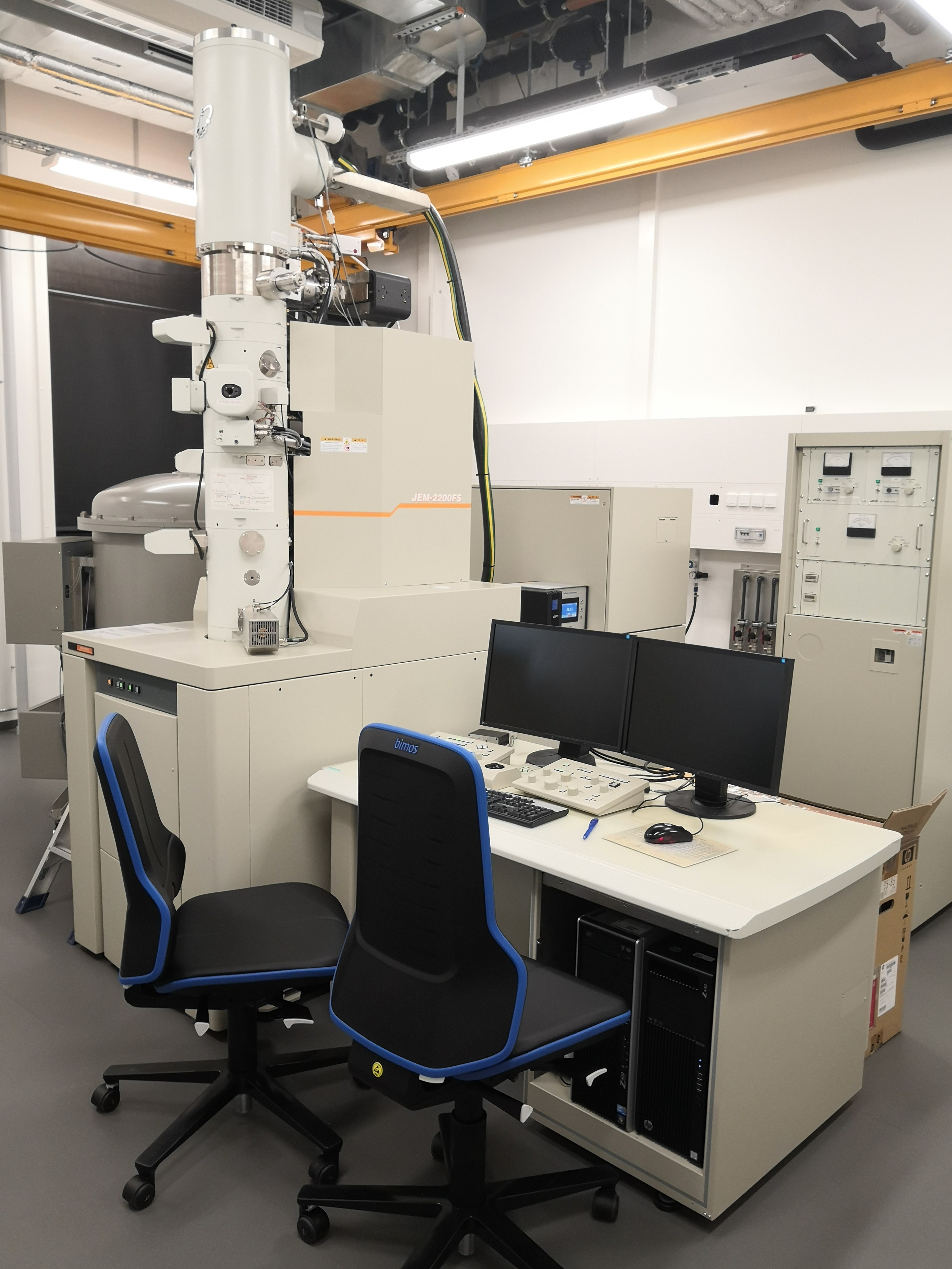
The instrument is a 200 kV JEM-FS2200 from JEOL with a field emission gun (FEG) and an on-line Omega Energy Filter allowing measurements at magnification from x 50 to x 1 M with a resolution of 0,2 nm in point and 0,1 nm in lattice. The Microscope is equipped with a Tietz CMOS camera with 2048 x 2048 pixels square area.
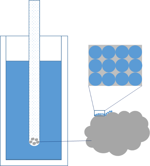
The soft matter samples have to be investigated either in dried or frozen state (Cryo-TEM) to be able to work in the necessary vacuum and to suppress blurring motion of the object as well as radiation damage.
As the samples have to be very thin (max~100nm), Cryo-TEM investigations require sophisticated sample preparation, the TEM laboratory comprises therefore an extended suite of preparation equipment. Users will be supported by JCNS scientists to conduct the suitable preparation and Cryo-TEM investigation.
Soft Matter investigation by room temperature TEM as well as Cryo-TEM on frozen thin specimen in liquid state and solid thin sections of polymers.
Detailed information is available at https://mlz-garching.de/tem
Field Emission Gun
200 kV acceleration voltage
Image recording on 2k by 2k CMOS Camera from Tietz Video Imaging and Processing System (TVIPS)
Image resolution around 0.17 nm in theory, but 2 nm for room-temperature TEM and 5 nm for Cryo-TEM due to the actual (temporary) environment
TEM sample can be moved plus/minus 1 mm in X and Y and tilted plus/minus 23° in X direction (Goniometer)
3 mm holey carbon coated cupper grids.
3 mm standard cupper grids. Gold grids on demand.
Thin liquid specimen, thin (ca. 100 nm) sections, eventually sample in powder state
Standard vacuum (10-6 Pa range) inside the instrument column
Specimen preparation devices for soft matter:
- Leica EM GP grid plunger for thin film of aqueous and organic solvent solution onto Cryogen (liquefied Ethane)
- Leica SCD050 for glow discharge to prepare the grid prior to freeze-plunging
- Leica UC-7/FC-7 Cryo-ultramicrotome using glass and diamond knife to perform ca. 100 nm thin cryo-sectioning on bulk polymer or resin embedded block specimen.
- Leica Freeze Fracture and Etching BAF060 to produce replicas from solution samples.
no
na
1
UAB
Spain
TEM JEOL 2011
na
Soft Matter investigation by Cryo-TEM on frozen thin specimen in liquid state.
XPS is a surface spectroscopic technique for quantitative measurements of the elemental composition or stoichiometry and the chemical state of the present elements, like their oxidation state and chemical bonds. XPS is highly surface sensitive, giving chemical and binding energy information from the a narrow region close to the surface.
In SEM a beam is scanned over a sample surface while a signal from secondary or back-scattered electrons is recorded. SEM is used to image an area of the sample with nanometric resolution, and also to measure its composition, crystallographic phase distribution and local texture.
SAXS is a non-destructive and versatile method to study the nanoscale structure of any type of material (solid, liquid, aerosols) ranging from new nanocomposites to biological macromolecules. Averaged particle sizes, shapes and distributions, porosity, degree of crystallinity and electron density maps with nanometer precision can be obtained.
PL is a non-contact, non-destructive method of probing the electronic structure of materials, often used in the context of semiconductor devices to determine the bandgap energy, the composition of heterostructures, the impurity levels, the crystal quality, and to investigate recombination mechanisms.
The Brunauer-Emmett-Teller method can be applied for determination of the specific surface area of a solid material. The volume specific surface area of a particulate material might be the basis of a decision whether it is considered to be nanomaterial according to the European Commission’s Recommendation on the definition of nanomaterial.