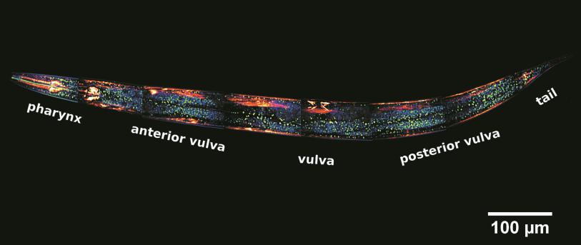
The facility provides access to state-of-the-art imaging infrastructure incorporating the latest technology in multiphoton microscopy and optical imaging, with a mission of enabling researchers to visualize and monitor fundamental processes deep inside molecular structures including soft materials, living cells and organisms. Sophisticated laser scanning microscopic instrumentation, ultra-sensitive digital cameras and specialized fluorescence probes make it possible to visualize cellular events in real time down to the molecular level. In addition, two-photon microscopy allows unparalleled detail in soft matter imaging and is extremely useful in monitoring cellular processes in vivo, enabling prolonged observations that are not possible with classic confocal microscopes.
The facility comprises of two laser scanning confocal microscope units and a spinning disk confocal microscopy system and a NLO/DUO/InTune multiphoton microscope offering capabilities, such as FRAP, FRET, FLIM, DICM, FC/DF, FLIP and live cell Imaging. In addition a Hybrid Photoacoustic and Laser Scanning Confocal Microscopy workstation is in operation offering real time structural and functional imaging of optically opaque samples including soft materials, live cells, tumor spheroids and biomimetic tissue samples.
In SEM a beam is scanned over a sample surface while a signal from secondary or back-scattered electrons is recorded. SEM is used to image an area of the sample with nanometric resolution, and also to measure its composition, crystallographic phase distribution and local texture.
In TEM/Scanning TEM (STEM) high energy electrons incident on ultra-thin samples, allow imaging, diffraction, electron energy loss spectroscopy and chemical analysis of solid materials with a spatial resolution on the order of 1-2 Å. Samples must have a thickness of a few tens of nanometres and are prepared in sample preparation laboratory.
AFM is a surface sensitive technique permitting to obtain a microscopic image of the topography of a material surface and certain properties (like friction force, magnetization properties…). Typical lateral image sizes are within a range of only a few Nanometers to several Micrometers, and height changes of less than a Nanometer.
Laser patterning is a technique for the controlled patterning of materials at micro- and nano-scales. It offers the ability to directly write patterns on the surface and complex 3D channels into the bulk of solid materials, also biomaterials. Applications can range from microfluidic systems and sensors to tissue engineering scaffolds.