Flow cytometry (FC) is a technique used to detect and measure physical and chemical characteristics of a population of cells or particles. In this process, a sample containing cells or particles is suspended in a fluid and injected into the flow cytometer instrument.
A flow cytometer has five main components: a flow cell, a measuring system, a detector, an amplification system, and a computer for analysis of the signals. In general, for the analysis, single-cell suspension must first be prepared.
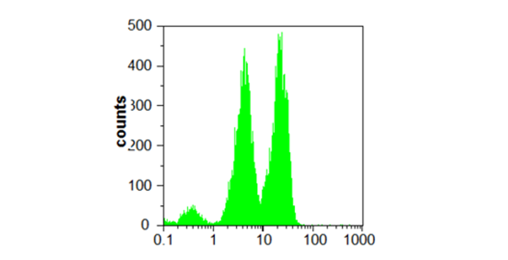
Our compact benchtop FC has two light sources can be used for applications in healthcare, microbiology, quality control or others. Absolute counting of cells, cellular subsets and other particles is performed in real-time on a volumetric basis.
The instrument is highly versatile and can be equipped for most applications in cell and particle analysis and absolute counting. Our instrument detects forward and side scatter signals using two laser lines (488 nm and UV).
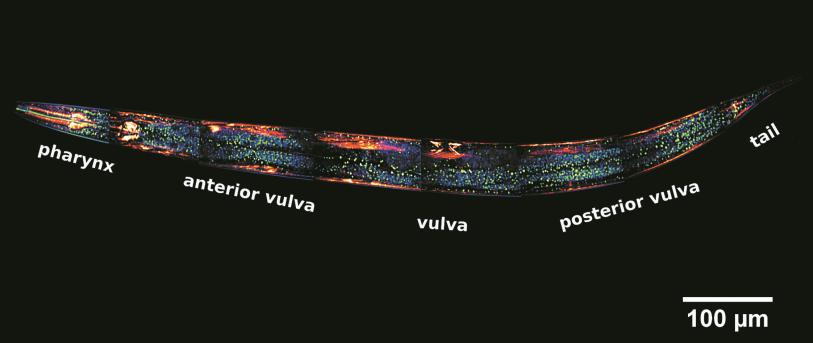
This technique has the mission of enabling researchers to visualise and to monitor cellular events in real time and in vivo down to the molecular level, enabling prolonged observations that are not possible with classic confocal microscopes and allowing unparalleled detail in soft matter imaging.
In TEM/Scanning TEM (STEM) high energy electrons incident on ultra-thin samples, allow imaging, diffraction, electron energy loss spectroscopy and chemical analysis of solid materials with a spatial resolution on the order of 1-2 Å. Samples must have a thickness of a few tens of nanometres and are prepared in sample preparation laboratory.
AFM is a surface sensitive technique permitting to obtain a microscopic image of the topography of a material surface and certain properties (like friction force, magnetization properties…). Typical lateral image sizes are within a range of only a few Nanometers to several Micrometers, and height changes of less than a Nanometer.
Fluorescence Correlation Spectroscopy (FCS) is a fluorescence based technique that gives information on the diffusion of fluorescent molecules or objects. From the diffusion times, the size of diffusing species can be determined, and the interaction among molecules or nanoparticles can be studied.
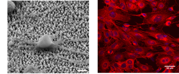
The Cell Culture Facility provides the necessary equipment (basic cell culture, sample preparation, functional assays and imaging equipment) for the study of the effect of biomaterials on specific cell behavior and function and cellular responses, such as cell survival, adhesion, morphology, proliferation, growth, migration and differentiation.