X-ray analysis
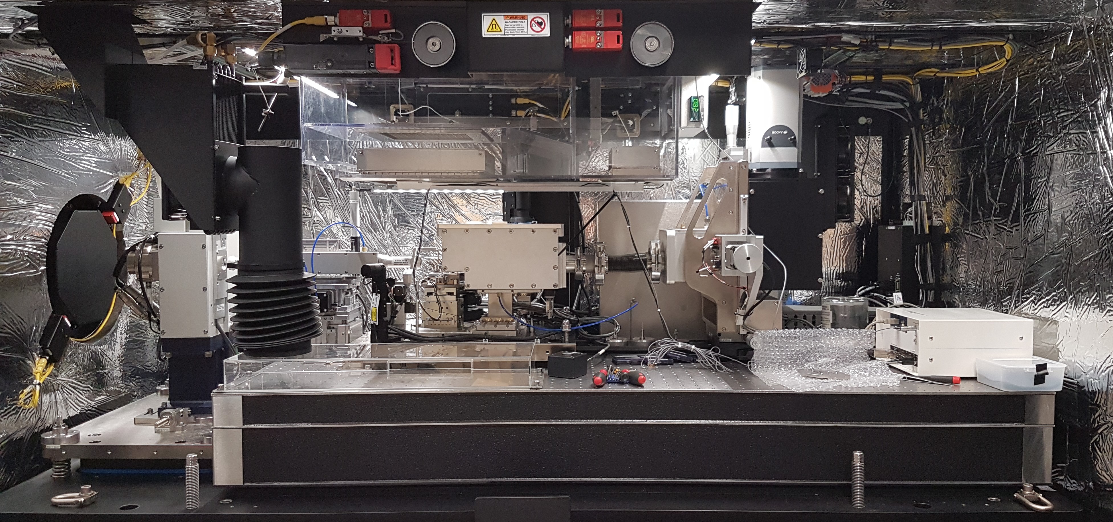
SAXS Small Angle X-ray Scattering
SAXS is a non-destructive and versatile method to study the nanoscale structure of any type of material (solid, liquid, aerosols) ranging from new nanocomposites to biological macromolecules. Averaged particle sizes, shapes and distributions, porosity, degree of crystallinity and electron density maps with nanometer precision can be obtained.
XRI X-Ray Imaging
NanoCT with in situ nanomechanical tests (high mechanical stability and a maximum force of 0.8 N) enables the non-destructive structural study under load, of the inner structure of 3D samples. Resolution down to 50 nm. It can be adjusted for absorption and phase contrast and can be operated with a large field of view or at high resolution.
XRT X-Ray Tomography
X-ray Computed Tomography (CT) is an indirect non-destructive technique for visualizing interior features within solid objects, and for obtaining digital information on their 3-D geometries and properties. Scanning x-ray tomography can also be performed using an x-ray beam (which size determines the resolution) focused on the sample.
XRR X-Ray Reflectivity
X-Ray (specular) Reflectivity or X-Ray Reflectometry, XRR, is a grazing incidence X-ray elastic scattering technique sensitive to surface electron densities. It is used to characterize flat and relatively smooth surfaces and layered samples proofing information about superficial layers: thickness, density, roughness, index of refraction.
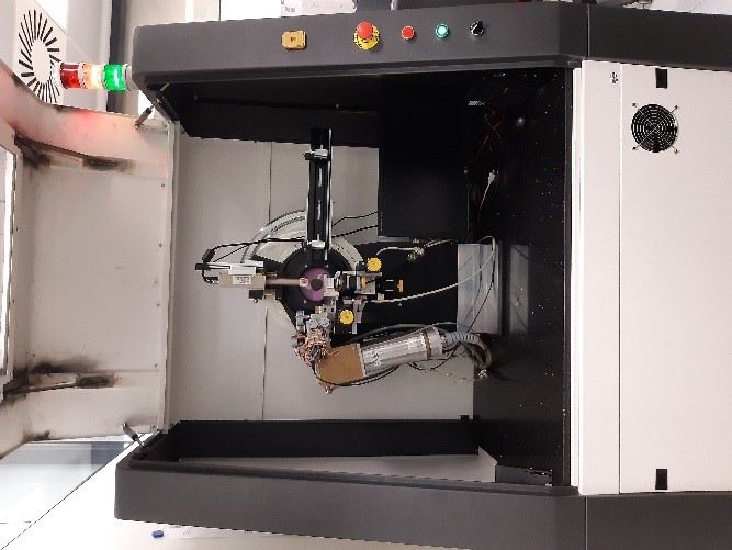
XRD X-Ray Diffraction
XRD provides non-destructive information on the structural order of a material. At large scattering angles XRD permits to identify different crystal phases and to quantify lattice distances and crystalline volume fractions. At low angles of incidence the surface roughness of a single crystal and the thickness of a deposition layer can be obtained.
XRM X-Ray Microscopy
Scanning Transmission X-Ray Microscopy (STXM) is a non-invasive x-ray microscopy technique that enables the acquisition of images with elemental, chemical, and magnetic sensitivity. Typical STXM images exhibit a spatial resolution on the order of 10-20 nm, depending on the zone plate employed to focus the x-rays, and a typical lateral image size


