X-Ray Microscopy
Structural & Morphology Characterization (X-ray analysis)
STXM is a non-invasive x-ray microscopy technique that can be employed for the space-, time- and spectroscopically-resolved investigation of micro- and nanostructured materials. In this technique, the sample under investigation is positioned in the focal spot of a focused x-ray beam, and the transmitted x-ray intensity is recorded with a suitable detector (typically, a photomultiplier tube or an avalanche photodiode). To acquire STXM images, the sample is raster scanned with a piezoelectric stage, and the transmitted intensity is recorded at each point of the scan.
Thanks to the use of x-rays as the probing mechanism, STXM allows easy access to a number of x-ray spectroscopic contrast mechanisms that allows imaging with quantitative sensitivity to not only the elemental composition, but also the chemical state of the sample material (i.e. oxidation state and/or molecular structure), with relatively low radiation damage. Furthermore, images with magnetic sensitivity can be acquired with STXM when illuminating the sample with circularly polarized x-rays, or with sensitivity to molecular orientation with linearly polarized x-rays.
Typical STXM images exhibit a spatial resolution on the order of 10-20 nm, depending on the Fresnel zone plate employed for the focusing of the x-rays. The lateral size for typical STXM images is on the order of 1-100 µm. Finally, if an avalanche photodiode is employed as detector, time-resolved STXM images with a temporal resolution down to 100 ps can also be acquired.

Instruments datasheets
Also consider
EUV-IL Extreme Ultra Violet - Interference lithography (temporarily unavailable)

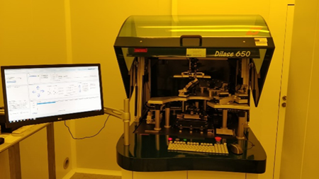
DWL Direct write lithography