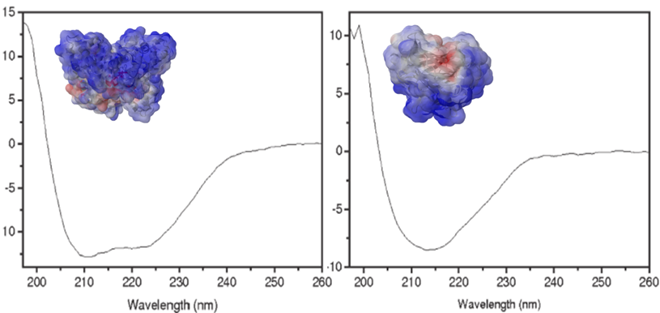
CD is an absorption spectroscopy method based on the differential absorption of left and right circularly polarized light. Optically active chiral molecules will preferentially absorb one direction of the circularly polarized light. The difference in absorption of the left and right circularly polarized light can be measured and quantified. UV CD is used to determine aspects of protein secondary structure.
In fact, alpha-helical and beta-sheet secondary structures of proteins have specific CD spectra in the 180-260 nm wavelength range, that are very different from unfolded proteins. Using well developed algorithms it is possible to deconvolute the CD spectra of a given protein to estimate the percentage of each secondary structure elements. By following the disappearance of secondary structure while increasing temperature it is possible to measure the thermal stability of the protein and to assess its relative stability in different buffers. CD can also be used to study how proteins change structure and/or stability when forming protein-nanoparticle complexes.
The technique is quite sensitive and requires limited amounts of material. It is essential that the buffer system (or other chemical compounds added to the protein sample) uses non chiral molecules, nor has strong absorption in the 190-300 nm wavelength.
The setup at CNR-IOM-Perugia allows for thermal cycling and multivariate analysis on demand to distinguish spectral features. This data analysis method is particularly useful for samples where intermediate states may occur during thermal unfolding or when external parameters are varied (he figure shows non-canonical DNA sequences as a function of temperature).
Circular dichroism (CD) is a spectroscopic technique that allows the rapid determination of the secondary structure and folding properties of purified proteins. The most widely used applications of protein CD are to determine whether an expressed, purified protein is folded and its thermal stability.
CNR-IOM (PG)
Italy
Circular Dichroism
Folding and binding of proteins, sugars, peptides, DNA/RNA nanosequences, oligomers
na
150W air-cooled Xenon lamp
Head-on photomultiplier tube
163 nm - 800 nm (JASCO-810)
Band width. Range: 0.01 nm – 15 nm
0.2 nm at 163 to 180 nm, 0.1 nm at 180 to 250nm, 0.3 nm at 250 to 500 nm, 0.8 nm at 500 to 800 nm
Liquid state - Materials: proteins, sugars, peptides, DNA/RNA nanosequences, oligomers - Concentrations: from nano to millimolar (around 0.01 mg/ml to 10 mg/ml)
Copper sample holder, temperature range from 20 °C to 100 °C - Rectangular quartz cell: 0.01 mm, 0.1 mm, 0.5 mm, 1 mm, 2 mm, 10 mm
*Set-up equipped with a collimated continuous LED source (ThorLabs) with wavelength of 430 nm and power of 500 mW for ex-situ preparation of irradiated materials. Samples are typically placed in a quartz cuvette at a distance of ~50 mm from the LED source where the collimated light spot has a diameter of ~25 mm. These conditions can be adjusted based on the experiments.
*Multivariate Analysis can be applied on request.
In SEM a beam is scanned over a sample surface while a signal from secondary or back-scattered electrons is recorded. SEM is used to image an area of the sample with nanometric resolution, and also to measure its composition, crystallographic phase distribution and local texture.
AFM is a surface sensitive technique permitting to obtain a microscopic image of the topography of a material surface and certain properties (like friction force, magnetization properties…). Typical lateral image sizes are within a range of only a few Nanometers to several Micrometers, and height changes of less than a Nanometer.