The microarray scanner measures the fluorescence intensity of labeled sample nucleic acid (DNA and RNA) bound to microarrays. Its ability to measure fluorescence from two dyes simultaneously facilitates all two-color microarray studies. This technology provides for rapid, high-quality, automated scanning of microarrays. The microarray scanner uses two lasers, a SHG-YAG laser (532 nm) and a helium-neon laser (633 nm). The lasers excite Cyanine-3 (Cy-3) and Cyanine-5 (Cy-5) labeled RNA or DNA to measure fluorescence after hybridization of the target nucleic acid to the microarray probes. The microarray scanner is optimized for high signal-to-noise performance in the Cy-3 (550—610 nm) and Cy-5 (650—750 nm) emission bands, with a wide dynamic range (up to five orders of magnitude) and low spectral cross-talk. This allows for measurement of a very broad range of target concentrations and for higher data confidence at lower signal levels. The laser excitation is scanned rapidly back and forth across the microarray. Fluorescence from the labeled samples is converted to an electrical signal by a high-performance PMT. Very low noise amplifiers and digital integrators process the PMT signal into a digital measurement that is recorded.
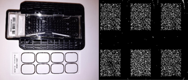
The microarray scanner is a laser-induced fluorescence scanner designed to read microarrays printed on standard 1 in × 3 in slides. The microarray scanner measures the fluorescence intensity of labelled sample nucleic acid (DNA and RNA) bound to microarrays.
JRC - ISPRA
Italy
Protein microarray scanner
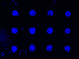
The microarray scanner measures the fluorescence intensity of labelled sample (nucleic acid, peptidic, proteic or any other type), bound to microarrays. Its ability to measure fluorescence from two dyes simultaneously facilitates all two-color microarray studies. This technology provides for rapid, high-quality, automated scanning of microarrays. The microarray scanner uses two lasers, with excitation wavelenght of 670 and 785 nm. The lasers excite compatible fluorophores (Alexa Fluor (700, 680), Dylight 680 or IRDye 680 on one hand and Alexa Fluor 790, Dylight 800 and IRDye 800) used to label target molecules after binding to the microarray probes. The microarray scanner is optimised for high sensitivity, with NIR lasers to avoid support background and real-time autofocus for a perfect homogeneity across the surface. The dynamic range of measurement allows to detect the lowest signals while avoiding saturation. Fluorescence from the labelled samples is detected in a real-time confocal manner with 2 photomultipliers. The image is acquired in tiff format by the software MAPIX, which also permits the spot quantification.
AFM is a surface sensitive technique permitting to obtain a microscopic image of the topography of a material surface and certain properties (like friction force, magnetization properties…). Typical lateral image sizes are within a range of only a few Nanometers to several Micrometers, and height changes of less than a Nanometer.
Non-contact printing is a technology based on a piezoelectric system, which consists in depositing drops of very low volume of samples (100 pL) without touching the surface, avoiding the risks of surface contamination. This technique is very flexible both in term of sample and of surface to modify.