Optical Thin Film Metrology

Structural & Morphology Characterization (Light and acoustic microscopy)

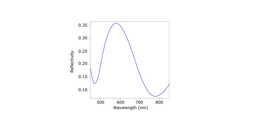
The principle of the thickness measurement is based on the interference patterns in the reflectivity spectrum of the layered sample. The measurement is performed without contact to the sample surface. Fast algorithms enable the performing pf the optical measurement and the calculation of the thickness with a high repetition rate and high reproducibility. In a typical configuration, the light reflected from the sample is collected by pick-up optics connected to the camera adapter of a microscope. An optical fiber guides the light to the spectrometer and the spectrum is detected by a high performance photo diode array. The measured spectrum from a layered sample shows typical interference effects which depend on the thickness and the optical properties of the materials. Therefore the film thickness can be determined if the optical properties and the sequence of films are known. Typical thicknesses between 50 nm and 20 μm can be measured with an accuracy of 1 nm. Examples of areas of applications include semiconductor technology (SiO2, Si3N4, resists etc), optical components, films on glass for liquid crystal displays, transparent coating on metals and other substrates etc.
i
@
          provided at NFFA-Europe laboratories by:
CSIC-CNM
Spain
FORTH
Greece
PSI
Switzerland
@
          provided by:
EURONANOLAB
France

Instruments datasheets

PSI
Switzerland
Optical thin film metrology @ Laboratory for Micro- and Nanotechnology
Optical thin film metrology
50 nm to 20 μm
450-920 nm spectral range
Photodiode array with 512 channels
Accuracy 1nm, precision (3σ) 0.1 nm
Thin films on various substrates
Clean room
EURONANOLAB
France
OTFM at EURONANOLAB - IEMN
EURONANOLAB
France
OTFM at EURONANOLAB - FEMTO-ST
EURONANOLAB
France
OTFM at EURONANOLAB - FBK
EURONANOLAB
France
OTFM at EURONANOLAB - IMM
i
@
          provided at NFFA-Europe laboratories by:
CSIC-CNM
Spain
FORTH
Greece
PSI
Switzerland
@
          provided by:
EURONANOLAB
France

Also consider

Structural & Morphology Characterization

SEM Scanning Electron Microscopy

In SEM a beam is scanned over a sample surface while a signal from secondary or back-scattered electrons is recorded. SEM is used to image an area of the sample with nanometric resolution, and also to measure its composition, crystallographic phase distribution and local texture.

Structural & Morphology Characterization

AFM Atomic Force Microscopy

AFM is a surface sensitive technique permitting to obtain a microscopic image of the topography of a material surface and certain properties (like friction force, magnetization properties…). Typical lateral image sizes are within a range of only a few Nanometers to several Micrometers, and height changes of less than a Nanometer.

Electronic & Chemical & Magnetic Characterization

RS Raman spectroscopy

Raman spectroscopy (RS) investigates the vibrational properties of a sample and provides chemical as well as structural information. RS does not require any specific sample preparation, size or condition and may be combined with micron/nano spatial resolution when operated using a confocal microscope/TERS or SNOM configuration.

Electronic & Chemical & Magnetic Characterization

Ellipsometry Ellipsometry

Ellipsometry is a contact-free, nondestructive method for characterization of the dielectric and optical properties (refractive index, absorption and thickness) of layered nanostructures in the size range of < 1 nm to several μm.