Nuclear Magnetic Resonance (NMR) is a non-destructive technique in which nuclear spin is manipulated to reveal molecular structure and dynamics information with atomic resolution. In the case of magnetic resonance imaging (MRI) and microscopy, a picture of the spin density can be generated with 3D pixel (voxel) dimensions down to ~ 10 µm in each direction. Nearly all elements have at least one isotope whose nucleus has non-zero spin (a quantum mechanical property, given the symbol I), and thus NMR can potentially probe a wide variety of materials in gas, liquid, solution, and solid phases.
Solid NMR can be used to retrieve information on phase structure in semicrystalline materials, to verify the extent of phase segregation in polymer blends, to measure molecular mobility at different length scales as a function of temperature, composition etc. It is also possible to study synthesis as well as functionalization reactions of polymers and/or inorganic substrates.
At KIT/ KNMF, our high field NMR system operates at 11.7 T, corresponding to a 1H resonance frequency of 500 MHz. One caveat to NMR / MRI is that it is a relatively insensitive technique. For reasonable measurement times, typical limit of detection values include: low µM concentration for small molecules (< 1500 Da), a few milligrams of material for large molecules in solution (e.g. proteins up to ~ 30 kDa), and several milligrams of material for solid samples.
At CNR the NMR Laboratory consists of two Spectrometers: Varian Mercury 400 MHz, equipped with a ID 1H-19F, 31P-15N bradband probe, and Agilent DD2 NMR System 500 MHz equipped with liquid (Agilent OneMNR), semisolid (Agilent Nanoprobe) and solid state (3.2 mm Agilent T3 triple resonance HXY MAS) probes.
Liquid state NMR spectroscopy;
(Semi)solid state NMR spectroscopy;
Magnetic resonance imaging; 2
Micro5 Microimaging probe (10 mm 1H, 5 mm 1H/13C);
HRMAS probe (4 mm, 1H/13C/2D);
BBFO liquid state probe (1H/19F - 31P-109Ag)
Liquid state samples - from 0.2 to 3 mL, temperature range -10 to 80 °C;
Semis-solid samples - from 1 to 100 mg, or from 0.04 - 0.1 mL, temperature range -10 to 80 °C
External magnetic field = 11.7 T
Magnetic resonance imaging capabilities. Sample must be confined in either 10 or 5 mm NMR glass sample tubes. Typical image resolution 50 micormeters isotropic possible within reasonable measurement time.
In TEM/Scanning TEM (STEM) high energy electrons incident on ultra-thin samples, allow imaging, diffraction, electron energy loss spectroscopy and chemical analysis of solid materials with a spatial resolution on the order of 1-2 Å. Samples must have a thickness of a few tens of nanometres and are prepared in sample preparation laboratory.
XPS is a surface spectroscopic technique for quantitative measurements of the elemental composition or stoichiometry and the chemical state of the present elements, like their oxidation state and chemical bonds. XPS is highly surface sensitive, giving chemical and binding energy information from the a narrow region close to the surface.
AFM is a surface sensitive technique permitting to obtain a microscopic image of the topography of a material surface and certain properties (like friction force, magnetization properties…). Typical lateral image sizes are within a range of only a few Nanometers to several Micrometers, and height changes of less than a Nanometer.
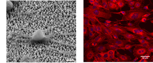
The Cell Culture Facility provides the necessary equipment (basic cell culture, sample preparation, functional assays and imaging equipment) for the study of the effect of biomaterials on specific cell behavior and function and cellular responses, such as cell survival, adhesion, morphology, proliferation, growth, migration and differentiation.
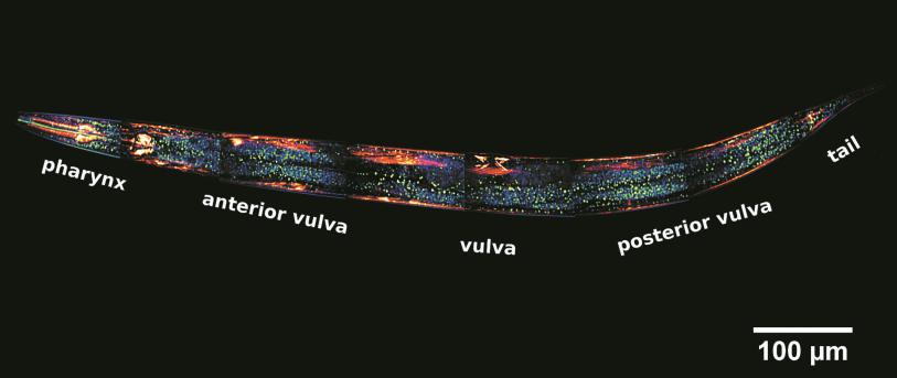
This technique has the mission of enabling researchers to visualise and to monitor cellular events in real time and in vivo down to the molecular level, enabling prolonged observations that are not possible with classic confocal microscopes and allowing unparalleled detail in soft matter imaging.