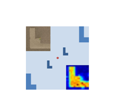
The offer comprises an advanced marker design tailored to specific requirements, the subsequent deposition of the markers by electron beam induced deposition (EBID), and a software tool that translates the position coordinates between the nano-science instruments via the marker system.
EBID markers are deposited using platinum (Pt) containing precursor material, making them suitable for localization by electron or X-ray excited Pt fluorescence, absorption, and their topography contrast. Markers are typically a few 10 mµ long, 15 µm wide and 0.1-1 µm thick and can be easily adapted to other specific need. The position coordinates are generated by a high-resolution field emission scanning electron microscope or an optical light microscope equipped with a translation stage and a digital readout of the lateral and height stage coordinates.
@
provided at NFFA-Europe laboratories by:
@
provided at NFFA-Europe laboratories by:
Also consider
Structural & Morphology Characterization
SEM Scanning Electron Microscopy
In SEM a beam is scanned over a sample surface while a signal from secondary or back-scattered electrons is recorded. SEM is used to image an area of the sample with nanometric resolution, and also to measure its composition, crystallographic phase distribution and local texture.
Structural & Morphology Characterization
AFM Atomic Force Microscopy
AFM is a surface sensitive technique permitting to obtain a microscopic image of the topography of a material surface and certain properties (like friction force, magnetization properties…). Typical lateral image sizes are within a range of only a few Nanometers to several Micrometers, and height changes of less than a Nanometer.
Structural & Morphology Characterization
XRD X-Ray Diffraction
XRD provides non-destructive information on the structural order of a material. At large scattering angles XRD permits to identify different crystal phases and to quantify lattice distances and crystalline volume fractions. At low angles of incidence the surface roughness of a single crystal and the thickness of a deposition layer can be obtained.
Structural & Morphology Characterization
SAXS Small Angle X-ray Scattering
SAXS is a non-destructive and versatile method to study the nanoscale structure of any type of material (solid, liquid, aerosols) ranging from new nanocomposites to biological macromolecules. Averaged particle sizes, shapes and distributions, porosity, degree of crystallinity and electron density maps with nanometer precision can be obtained.


