Digital holography
Nano to Micro/Macro (in vitro assays and cell analysis)
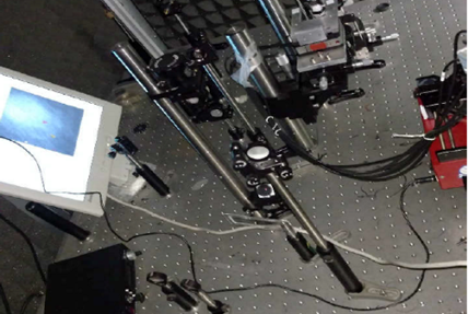
Digital Holography setups combine into a single platform the high-quality imaging provided by brightfield transmission optical microscopy with high numerical aperture objectives (water and oil immersion NA > 1.2), whole object wave front recovery provided by holography and numerical processing capabilities provided by custom developed software. The holographic architecture is implemented at the image space dividing the beam after the microscope lens, thus reducing the sensitivity of the system to vibrations and/or thermal changes in comparison to regular interferometers. Because of the off-axis holographic recording principle, quantitative phase images of live biosamples can be recorded in a single camera snapshot at full-field geometry without any moving parts. The use of water/oil-immersion imaging lenses maximizes the achievable resolution limit. This dual-mode microscope platform can be applied to the characterization (e.g. measuring cell height, volume, dry mass) of fixed or flowing living cells without additional markers.
The coherent illumination is aligned with the incoherent one following the same optical path until adichroic mirror DM2, where it becomes transmitted, allowing the assembly of a home-built Mach–Zehnder interferometric architecture. Concerning the numerical processing for retrieving the complex amplitude distribution of the sample in the DHM imaging path, a method based on spatial filtering at the Fourier plane from the recorded off-axis holograms is implemented.


