In Vitro Assays

Nano to Micro/Macro (in vitro assays and cell analysis)

Cytotoxicity is one of the most important indicators for biological evaluation in vitro studies. Cytotoxicity and cell viability assays are based on various cell functions. A broad spectrum of cytotoxicity assays is currently used in the fields of toxicology and pharmacology. There are different classifications for these assays:

(i) dye exclusion assays (Trypan blue, eosin, Congo red, …). Determination of membrane integrity is possible via dye exclusion method

(ii) colorimetric assays (MTT assay, MTS assay, XTT assay, WST-1 assay, WST-8 assay, LDH assay, …)

(iii) fluorometric assays (alamar Blue assay): Fluorometric assays of cell viability and cytotoxicity are easy to perform with the use of a fluorescence microscope, fluorometer, fluorescence microplate reader or flow cytometer.

(iv) luminometric assays (ATP assay). Luminometric assays provide fast and simple determination of cell proliferation and cytotoxicity in mammalian cells. These assays can be performed in a convenient 96-well and 384-well microtiter plate format and detection by luminometric microplate reader. When cells damaged lethally and lose membrane integrity, they lose the ability to synthetize ATP and the ATP level of cells decreases dramatically .

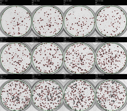
(v) Colony Forming Efficiency (CFE) is a clonogenic assay that measures the ability of a single cell to form a colony: CFE is a labelled free assay and usually more sensitive than the conventional biochemical methods probably because this test does not measure a specific biological effect, but rather cell death in general.

i
@
          provided at NFFA-Europe laboratories by:
CSIC-ICMAB
Spain
JRC - ISPRA
Italy
i
@
          provided at NFFA-Europe laboratories by:
CSIC-ICMAB
Spain
JRC - ISPRA
Italy

Also consider

Electronic & Chemical & Magnetic Characterization

XPS X-ray Photoelectron Spectroscopy

XPS is a surface spectroscopic technique for quantitative measurements of the elemental composition or stoichiometry and the chemical state of the present elements, like their oxidation state and chemical bonds. XPS is highly surface sensitive, giving chemical and binding energy information from the a narrow region close to the surface.

Structural & Morphology Characterization

TEM Transmission Electron Microscopy

In TEM/Scanning TEM (STEM) high energy electrons incident on ultra-thin samples, allow imaging, diffraction, electron energy loss spectroscopy and chemical analysis of solid materials with a spatial resolution on the order of 1-2 Å. Samples must have a thickness of a few tens of nanometres and are prepared in sample preparation laboratory.

Electronic & Chemical & Magnetic Characterization

ICP-MS ICP Mass spectroscopy (ICP-MS) (single particle analysis, trace element analysis)

ICP-MS allows measuring elements at trace levels. Dissolved sample is introduced into an argon plasma where the molecules are dissociates and further ionized. Singly-charged ions thus formed are directed into the mass spectrometer. A special application of ICP-MS (spICP-MS) allows measuring size and number concentration of a particle suspension.

Structural & Morphology Characterization

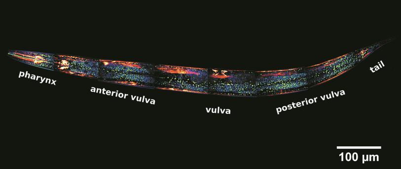
LSCM laser scanning confocal microscopy

This technique has the mission of enabling researchers to visualise and to monitor cellular events in real time and in vivo down to the molecular level, enabling prolonged observations that are not possible with classic confocal microscopes and allowing unparalleled detail in soft matter imaging.

Structural & Morphology Characterization

Cryo-TEM Cryo-Transmission Electron Microscopy

Cryo-Transmission electron microscopy is a technique for soft matter studies allowing real space investigations about shape and size distribution of particles, self-assembly and aggregation. Liquid sample can be directly observed on grid after blotting. An automatic plunge freezer is used to make cryo-specimen for direct imaging (Cryo-DI).