The BET technique uses adsorption of an inert gas (in our case nitrogen) to measure the outer and inner surface area of a solid sample. The sample has to be heat and vacuum pre-treated to remove the adsorbed molecules (mainly water) that might occupy the surface. After the pre-treatment procedure the sample is transferred to the measurement position. As the inert nitrogen and solids interact weakly, the solid material is cooled to the temperature of liquid nitrogen and kept under isothermal conditions during the whole measurement. The pressure of the nitrogen gas is increased gradually and a monolayer of adsorbed nitrogen molecules is built on the solid surface. The number of gas molecules in the formed monolayer is determined from the adsorbed gas volume. The “footprint” of the nitrogen molecule on the surface is known and the total surface area of sample can be calculated by using the BET equation. The mass specific surface area [m2/g] is derived from this total surface by dividing it with the mass of the sample. Multiplying this value with the skeletal density of the material [g/cm3] provides the volume specific surface area of the material.
Nitrogen adsorption measurements can provide information not only on the external surface area but also on the porosity of samples. Specific surface area affects functional properties of many different type of materials, including pharmaceuticals, catalysts, structural components.
The Brunauer-Emmett-Teller method can be applied for determination of the specific surface area of a solid material.
The BET technique uses adsorption of an inert gas (in our case nitrogen) to measure the outer and inner surface area of a solid sample.Nitrogen adsorption measurements can provide information not only on the external surface area but also on the porosity of samples. Specific surface area affects functional properties of many different type of materials, including pharmaceuticals, catalysts, structural components.
XPS is a surface spectroscopic technique for quantitative measurements of the elemental composition or stoichiometry and the chemical state of the present elements, like their oxidation state and chemical bonds. XPS is highly surface sensitive, giving chemical and binding energy information from the a narrow region close to the surface.
In SEM a beam is scanned over a sample surface while a signal from secondary or back-scattered electrons is recorded. SEM is used to image an area of the sample with nanometric resolution, and also to measure its composition, crystallographic phase distribution and local texture.
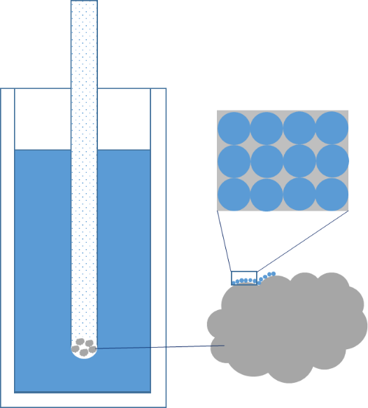
Cryo-Transmission electron microscopy is a technique for soft matter studies allowing real space investigations about shape and size distribution of particles, self-assembly and aggregation. Liquid sample can be directly observed on grid after blotting. An automatic plunge freezer is used to make cryo-specimen for direct imaging (Cryo-DI).
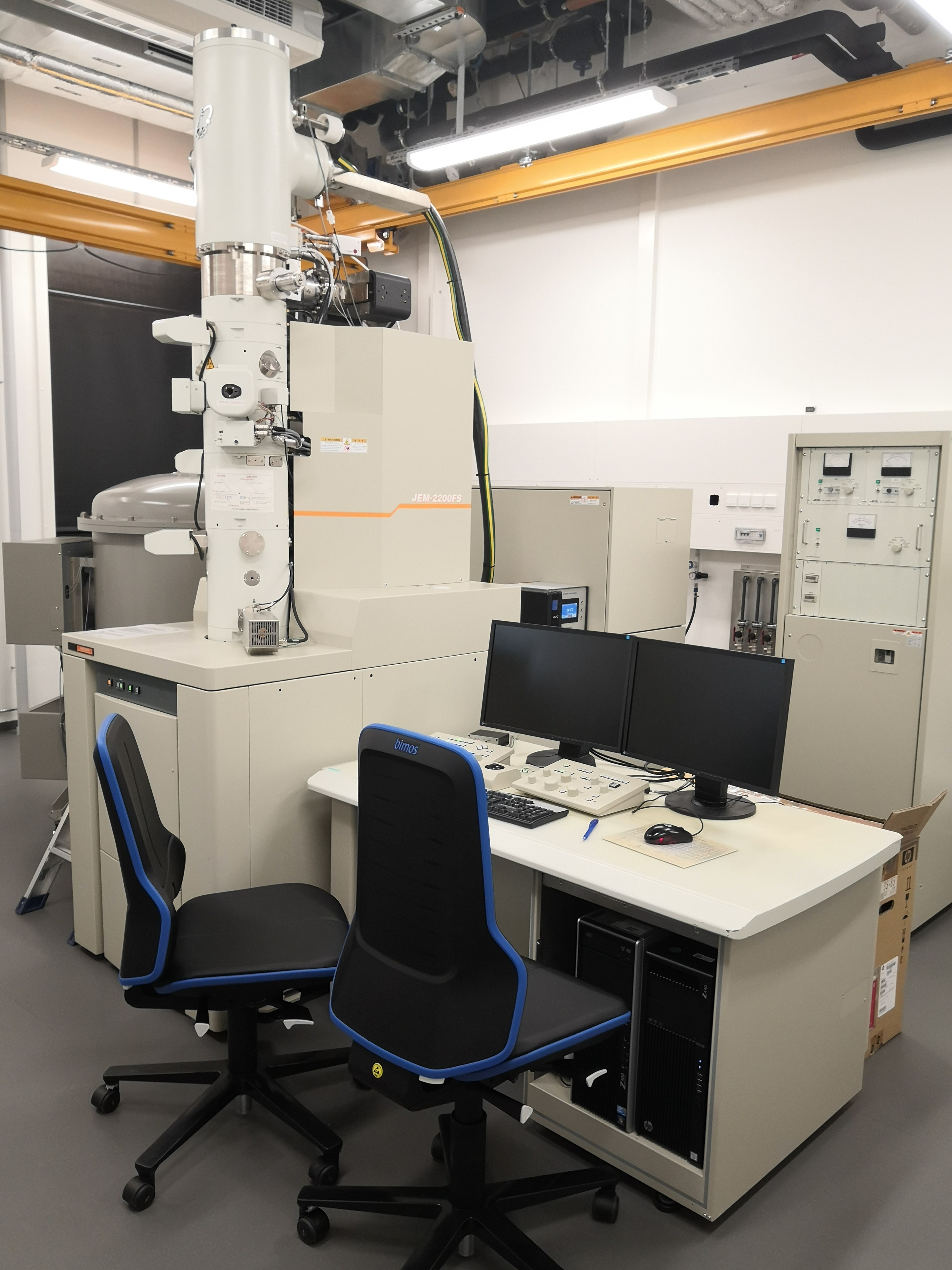
In TEM/Scanning TEM (STEM) high energy electrons incident on ultra-thin samples, allow imaging, diffraction, electron energy loss spectroscopy and chemical analysis of solid materials with a spatial resolution on the order of 1-2 Å. Samples must have a thickness of a few tens of nanometres and are prepared in sample preparation laboratory.
XRD provides non-destructive information on the structural order of a material. At large scattering angles XRD permits to identify different crystal phases and to quantify lattice distances and crystalline volume fractions. At low angles of incidence the surface roughness of a single crystal and the thickness of a deposition layer can be obtained.