Asymmetric Field Flow Fractionation with different detectors (MALS, DLS, UV-vis). Nanoparticle separation by sizes

Structural & Morphology Characterization (Dispersed-phases characterisation)

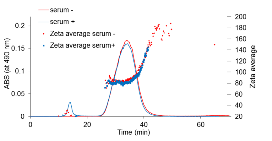
In this instrument, a thin, flat channel is sandwiched between two walls, one of which is solid and the other which is made from a membrane through which water can be pumped. When a liquid containing different sizes of nanoparticles passes through the channel, careful control of the liquid flows along the channel and across the membrane can be used to separate the particles depending on their size, usually with smaller particles exiting the channel first. Size measurement of the separated fractions using on-line coupled DLS or MALS detectors allows a more precise determination of particle size compared to batch mode light scattering based methods by avoiding the misleading effect of strongly scattering, large particles. The typical working range is <500 nm (steric elution range, where larger particles start to travel faster and elute first from the column). The instrument usually performs well for negatively charged and PEG coated nanoparticles or in the presence of detergents or stabilisers that help to minimise the interaction with the semi-permeable membrane wall of the channel. Fraction collector allows the analysis of separated size fractions by methods like TEM that are not compatible with direct on-line coupling

i
@
          provided at NFFA-Europe laboratories by:
JRC - ISPRA
Italy

Instruments datasheets

JRC - ISPRA
Italy
ASYMMETRICAL FIELD FLOW FIELD FRACTIONATION
The AF4 instrument separates nanoparticles according to their size in a liquid flow. The instrument can be on-line coupled with various concentration (UV-Vis, RI) and size measurement (DLS, MALS) detectors as well as with a fraction collector.
i
@
          provided at NFFA-Europe laboratories by:
JRC - ISPRA
Italy

Also consider

Structural & Morphology Characterization

TEM Transmission Electron Microscopy

In TEM/Scanning TEM (STEM) high energy electrons incident on ultra-thin samples, allow imaging, diffraction, electron energy loss spectroscopy and chemical analysis of solid materials with a spatial resolution on the order of 1-2 Å. Samples must have a thickness of a few tens of nanometres and are prepared in sample preparation laboratory.

Electronic & Chemical & Magnetic Characterization

ICP-MS ICP Mass spectroscopy (ICP-MS) (single particle analysis, trace element analysis)

ICP-MS allows measuring elements at trace levels. Dissolved sample is introduced into an argon plasma where the molecules are dissociates and further ionized. Singly-charged ions thus formed are directed into the mass spectrometer. A special application of ICP-MS (spICP-MS) allows measuring size and number concentration of a particle suspension.

Structural & Morphology Characterization

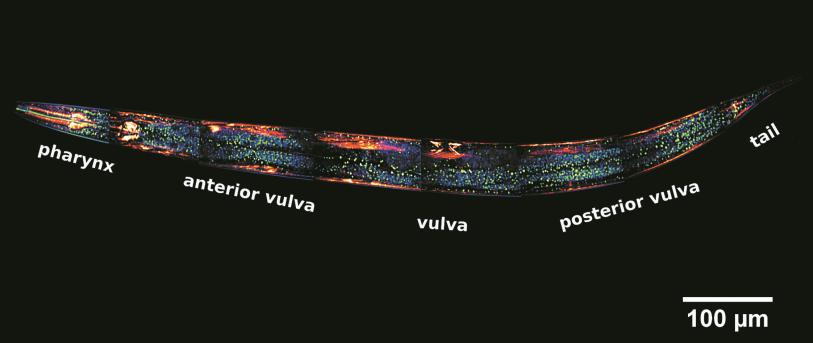
LSCM laser scanning confocal microscopy
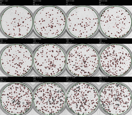
This technique has the mission of enabling researchers to visualise and to monitor cellular events in real time and in vivo down to the molecular level, enabling prolonged observations that are not possible with classic confocal microscopes and allowing unparalleled detail in soft matter imaging.

Nano to Micro/Macro

INA In Vitro Assays

In vitro assays on cell cultures allow the study of biological functions and phenomena: each type of assay corresponds to a biological function or mechanism. These assays are used to assess the safety of advanced materials and in particular nanomaterials.