Graphene-nanohydroxyapatite bone lamellar scaffold has mechanical strength that is comparable to natural bone.
Growth of mechanically strong and macroporous scaffolds is essential to future bioengineering. This work demonstrates the possibility of the synthesis of a new graphene-nanohydroxyapatite structure with high mechanical strength that is favorable for bone regeneration. Graphene-nanohydroxyapatite was synthesized by the hydrothermal method. Elongated nanohydroxyapatite crystals were grown on the layers of graphene with nanowindows, serving as active sites for crystalline growth.
Mechanical properties of the graphene-nanohydroxyapatite were studied on the dry scaffolds filled into the Teflon molds. The compressive strain of the graphene-nanohydroxyapatite reached ~13 MPa, being significantly higher than that of the pure nanohydroxyapatite due to the exceptional mechanical strength of graphene. The high mechanical strength appears due to firm contacts between graphene and nanohydroxyapatite. The molecular dynamics simulations show that the nanohydroxyapatite has a tendency to grow near the surface of nanowindows because of the electrostatic attractions between calcium from hydroxyapatite and oxygen groups on the surface of the nanowindows. The microstructure of the graphene-nanohydroxyapatite shows the presence of large macropores of nearly 100 nm in size, being available for the cell penetration and growth of the new bone structure.
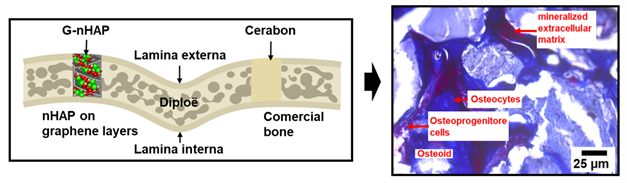
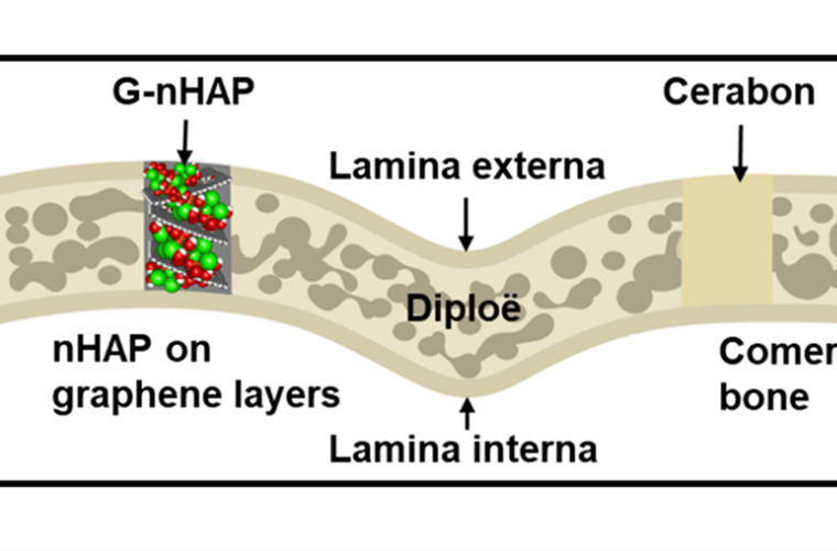
Graphene-nanohydroxyapatite (G-nHAP) filling the skull bone in rats with cerabon as a reference, showing the scaffold lamellar structure, osteoid cells and mineralized extracellular matrix after six weeks.
The graphene-nanohydroxyapatite has a paste-like consistency and can be used in dry and wet states reversibly, allowing easy transport, preservation, and embedding into bone defects. Graphene-nanohydroxyapatite can fill the bone defects as we demonstrated through in vivo experiments on Wistar rats. The paste-like material was filled into bone defects, and after six weeks we detected the cells responsible for the new bone formation. A higher volume density of the cells (osteocytes and osteoblasts) in graphene-nanohydroxyapatite than that in pure nanohydroxyapatite demonstrates that the cells have an affinity to grow near the hydrophilic surfaces of graphene with nanowindows.
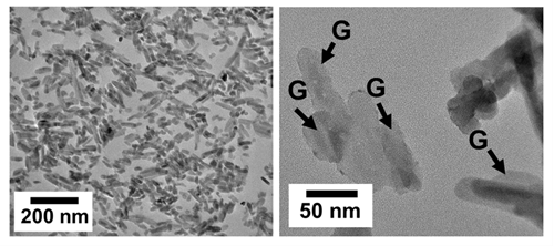
Transmission electron micrographs of the graphene nanohydroxyapatite, showing needle-like structures of nanohydroxyapatite in contact with graphene (G shown by arrows).
NFFA-Europe offered us a unique opportunity to conduct detailed characterizations of the graphene-nanohydroxyapatite scaffold including the transmission and scanning electron microscopies, thermogravimetry, Raman spectroscopy, and X-ray diffraction analysis. These characterizations contributed to an in-depth understanding of the scaffold structure, leading to successful scaffold preparation.
Kukobat, R.; Škrbić, R.; Jovičić, S.; Matičić, M.; Stojmenovski, A.; Bulajić, M.; Marin, S.; Bodroža, D.; Stević, D.; Atlagić, S. G.; Ignjatović, N.; Vallejos-Burgos, F.; Mercadelli, E.; Ponti, J.; Fumagalli, F.; Valsesia, A. Graphene Nanowindows as a Basis for Creating Mechanically Robust Nanohydroxyapatite Bone Lamellar Scaffolds. Carbon 2025, 243, 120589. https://doi.org/10.1016/j.carbon.2025.120589.
NFFA-Europe facilities from JRC – ISPRA and CNR-DSCTM were used for the bone scaffold characterization and analysis.
SEM and TEM observations were conducted to examine the surface morphology and microstructures of the graphene-nanohydroxyapatite scaffolds. The scaffolds with different contents of graphene were examined, showing their shape and morphology. TEM served for the cross-sectional observations of the scaffolds that were previously molded into silicon resin. Clear observations of the microstructures and large pores were obtained, contributing to a better understanding of our scaffolds.
Thermogravimetric analysis was used to examine the thermal stability of the scaffolds in nitrogen and air atmospheres. Raman spectroscopy served for the structural characterization of graphene before and after crystalline growth. The presence of the bands from defects and crystalline phases contributed to a deeper understanding of the interactions between graphene and nanohydroxyapatite.
XRD gave clear evidence of crystalline growth and the structure of nanohydroxyapatite. The presence of the hexagonal crystalline structure was evidenced, and the defects at different crystalline faces were estimated using the Scherrer equation. The differences in the crystalline sizes on different crystallographic faces suggest an interference of graphene with the crystalline growth of nanohydroxyapatite.

Dr. Radovan Kukobat completed his PhD in Materials Science at Shinshu University in 2017 for his work on the development of the Zn/Al complex dispersant for aqueous dispersions of single wall carbon nanotubes and the study of electrical and optical properties of the films derived from aqueous dispersions. He also conducted his postdoctoral research at Shinshu University on gas separation membranes. He worked on graphene-zeolite membranes for hydrogen separations, developing highly selective membranes with ultrahigh hydrogen permeability. He started his independent research at the University of Banja Luka in 2021, continuing to work on membranes for carbon-dioxide separation in addition to challenging the new area in biomedical engineering. His work in the biomedical field resulted in several publications, including drug adsorption/dissolution and bone scaffolds. He is actively searching for new routes for material synthesis with superior optical, electrical and surface properties for energy, environment, and bioengineering.

