Structural & Morphology Characterization
Dispersed-phases characterisation View all
MALS Multi Angle Light Scattering (MALS) (nanoparticle sizing)
MALS is a technique for measuring light scattered by a sample into a plurality of angles. It is used to determine both the absolute molar mass and the average size of molecules in solution. MALS is typically used as a flow-mode detector coupled to Asymmetric Flow Field Flow fractionation (AF4) or Size Exclusion Chromatography (SEC).
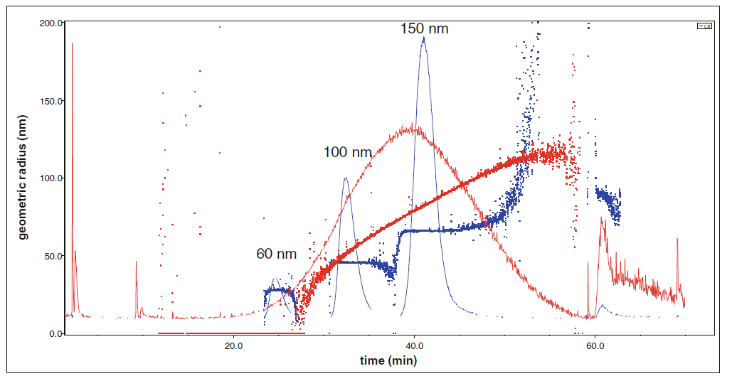
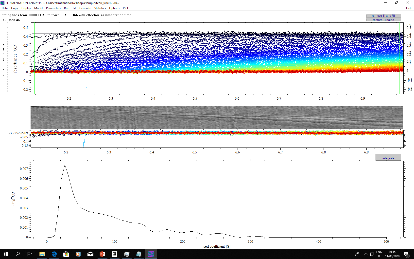
DCS Disk centrifuge sedimentation (nanoparticle sizing)
Disc Centrifuge Sedimemtation (DCS) is able to separate particles or agglomerates of particles according to their mass (size and density) by sedimentation in a liquid density gradient. The size of the particles is calculated from their sedimentation time.
DLS Dynamic Light Scattering
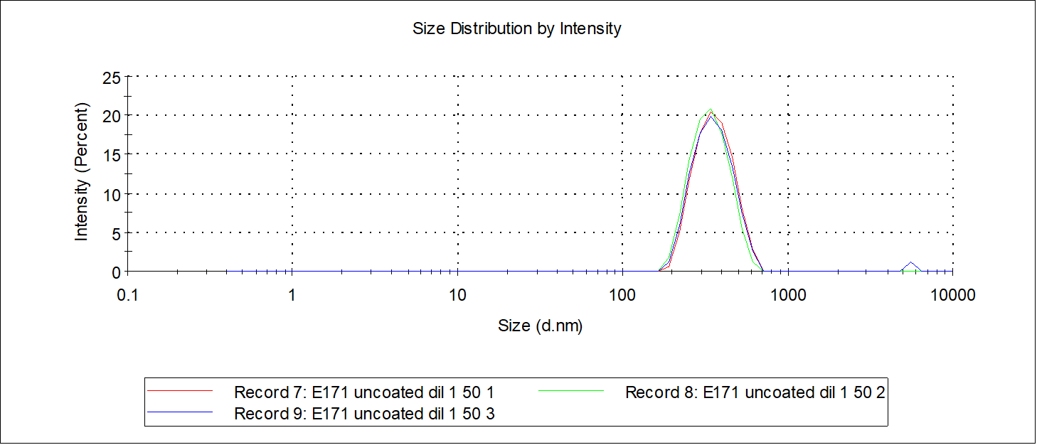
DLS is a powerful tool for investigating the diffusion behaviour of macromolecules or particles in suspension polar or non-polar liquid media. It gives an estimate of the size of the particles by means of mathematical relations between light scattering and diffusion behaviour of particles.
ζ-potential ζ-potential
This electroacoustic-based technology allows the measurement of the zeta potential of particles dispersed in liquid media that is strictly related with the colloidal stability of the suspensions.
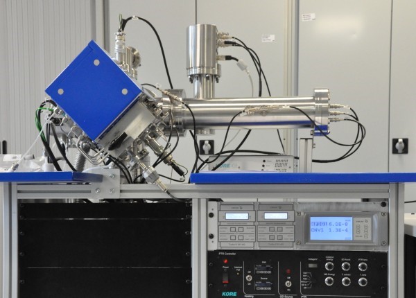
UAC Analytical Ultra Centrifuge (UAC) (nanoparticle/protein sizing)
The AUC is used to measure the size distribution of nanoparticles and macromolecules in suspension. The instrument performs well in the size distribution measurement of small nanoparticles from proteins and other macromolecules to viruses, liposomes and inorganic submicron particles.
WAS Wide Angle Static and Laser Diffraction
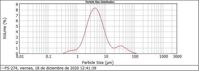
Laser diffraction (LD) measures particle size distribution in many systems (e.g. powders, suspensions, emulsions) in the range 0,1 µm - 3 mm. The intensity data is analyzed to calculate the size of the particles, using the Mie theory of light scattering. The particle size is reported as a volume equivalent sphere diameter.
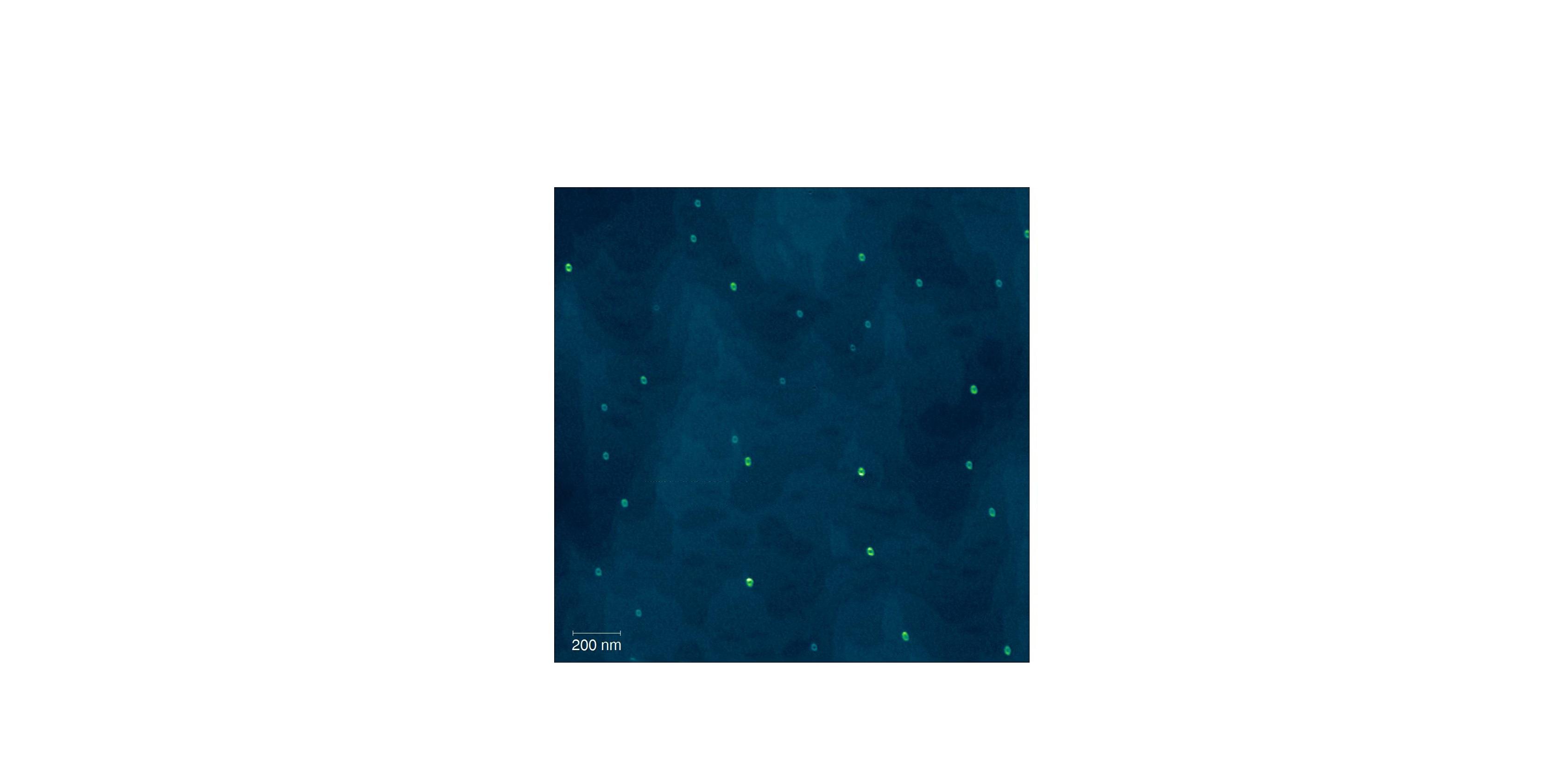
NPTA Nanoparticle Tracking Analysis
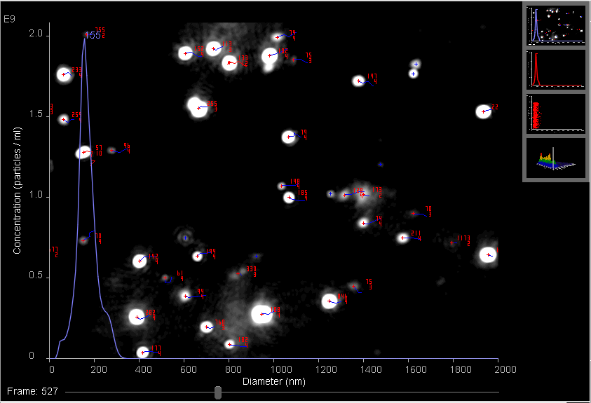
The NPTA technique allows the determination of the particle size distribution (PSD) profile and concentration of particles with a diameter of approximately 10-1000 nanometers in liquid suspension. Moreover, the fluorescence mode provides specific results for labelled particles.
AFFF Asymmetric Field Flow Fractionation with different detectors (MALS, DLS, UV-vis). Nanoparticle separation by sizes
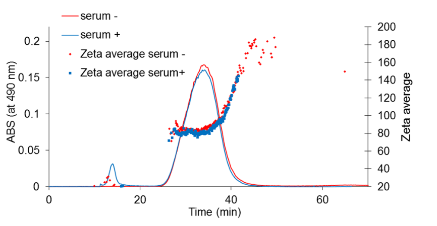
The AF4 instrument separates nanoparticles according to their size in a liquid flow. The instrument can be on-line coupled with various concentration (UV-Vis, RI) and size measurement (DLS, MALS) detectors as well as with a fraction collector.
Scanning probe microscopy View all
STM Scanning Tunneling Microscopy
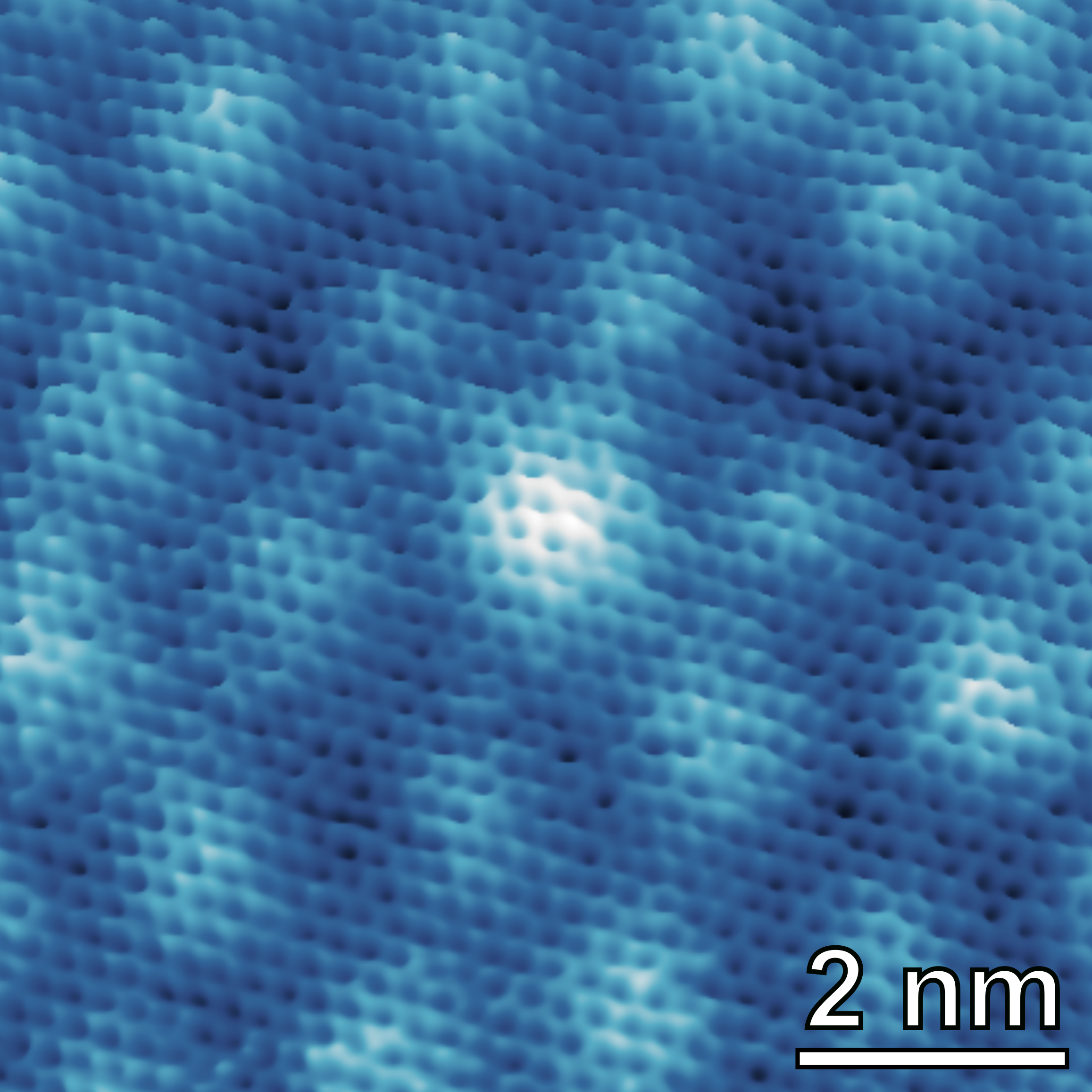
STM allows imaging conductive surfaces at the atomic scale. It is possible to characterize the distribution of surface terraces and steps, as well as to determine the atomic arrangement of (ordered) surface (over)structures.
AFM Atomic Force Microscopy
AFM is a surface sensitive technique permitting to obtain a microscopic image of the topography of a material surface and certain properties (like friction force, magnetization properties…). Typical lateral image sizes are within a range of only a few Nanometers to several Micrometers, and height changes of less than a Nanometer.
Electron and ion beam technologies View all
IBA IBA (Ion Beam Analysis)
IBA consists of bombarding samples with few MeV ions to obtain information about depth profile and absolute element concentration (at/cm²) from a few nm up to a few µm. It combines different techniques that have their own features (light/heavy/trace element detection, etc.) but can be combined to work in a synergic way.
SIMS Secondary Ion Mass Spectrometry
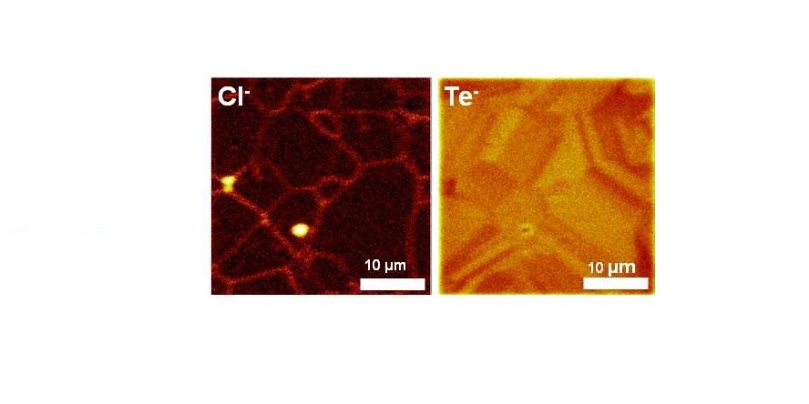
By SIMS, samples are bombarded; sputtered and ionized atoms accelerate towards a mass spectrometer, providing surface mass spectra, images with lateral resolution in the 0.1 to 10 µm range and depth profiles with depth resolution in the 1 to 10 nm range. SIMS can be quantitative up to ppb sensitivity when reference samples are used.
FIB prep for TEM FIB sample preparation for TEM
In FIB an energetic beam of ions creates local removal of material by sputter erosion. Combination with an electron column, allows 2D and 3D nanometric structures to be achieved. Characterization by different detectors (SEM, BSE, STEM, X-Ray) is possible. FIB is the preferred technique to prepare extremely thin slices for TEM investigations.
APT Atom Probe Tomography
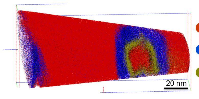
A voltage and/or a laser pulse is used to field evaporate atoms from the end of a specimen in the form of a sharp tip. By measuring the time-of-flight and the position of the evaporated atoms on a position sensitive detector a 3-D reconstruction of the position and chemical nature of detected atoms is possible with sub-nm resolution.
SEM Scanning Electron Microscopy
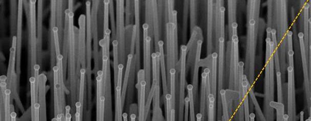
In SEM a beam is scanned over a sample surface while a signal from secondary or back-scattered electrons is recorded. SEM is used to image an area of the sample with nanometric resolution, and also to measure its composition, crystallographic phase distribution and local texture.
TEM Transmission Electron Microscopy
In TEM/Scanning TEM (STEM) high energy electrons incident on ultra-thin samples, allow imaging, diffraction, electron energy loss spectroscopy and chemical analysis of solid materials with a spatial resolution on the order of 1-2 Å. Samples must have a thickness of a few tens of nanometres and are prepared in sample preparation laboratory.
Cryo-TEM Cryo-Transmission Electron Microscopy
Cryo-Transmission electron microscopy is a technique for soft matter studies allowing real space investigations about shape and size distribution of particles, self-assembly and aggregation. Liquid sample can be directly observed on grid after blotting. An automatic plunge freezer is used to make cryo-specimen for direct imaging (Cryo-DI).
CLEM Correlative light-electron microscopy
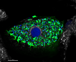
The Correlative Light and Electron Microscopy technique (CLEM) is a powerful approach that enables comprehensive investigations by combining optical microscopy, – using fluorochromes for biomolecule localization – and transmission electron microscopy (TEM), which provides high-resolution images at the nanoscale.
Light and acoustic microscopy View all
FM Hybrid tomographic and Light Sheet Fluorescence Microscopy
The hybrid Optical Projection Tomography and Selective Plane Illumination Microscopy (OPT-SPIM) is used to produce optical sectioning of molecular structures (soft materials, biological tissue, etc) with submicron resolution.
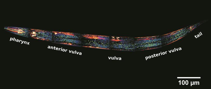
NLM Nonlinear microscopy
NLM takes advantage of tightly focused ultra-short laser pulses (fs) to excite non-linear optical phenomena like Second Harmonic Generation, Third Harmonic Generation, Two-Photon and Three-photon excited Fluorescence. Along with laser raster-scanning of the sample, non-linear imaging microscopy of large areas is accomplished within seconds.
OAM Optoacoustic microscopy
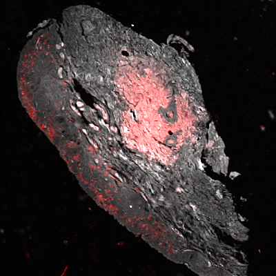
Photoacoustic microscopy is a novel diagnostic technique, mainly developed in biomedical research. It offers label-free optical absorption contrast of several biological molecules. Combination with traditional confocal fluorescence microscopy may provide multiparametric information high specificity, sensitivity and spatial resolution.
CM Confocal microscopy
Confocal microscopy (CM) is an optical imaging technique that creates a virtual plane or slice, many micrometers deep within the analyzed sample. Compared to conventional microscopy, it provides fine detailed images of higher quality and with more contrast. In addition, virtual 3-D images of the analyzed microstructure can be obtained.
LSCM laser scanning confocal microscopy
This technique has the mission of enabling researchers to visualise and to monitor cellular events in real time and in vivo down to the molecular level, enabling prolonged observations that are not possible with classic confocal microscopes and allowing unparalleled detail in soft matter imaging.
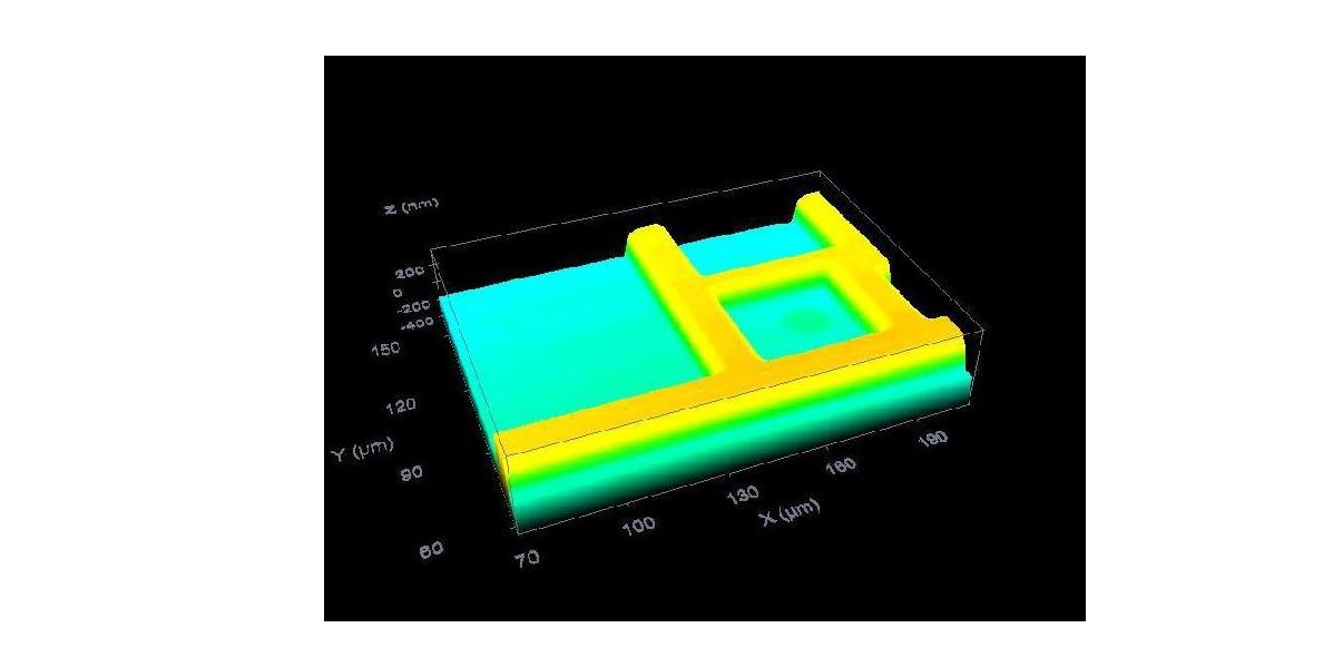
OTFM Optical Thin Film Metrology
Thin film reflectometry is a fast and easy technique used to measure the thickness of thin transparent and semi-transparent films on transparent and absorbing substrates. It covers a broad thickness range and can be applied to a high variety of material systems due to the large and extensible material library.
Neutron characterisation View all
NI** Neutron Imaging (temporarily unavailable)
Neutron imaging allows performing radiography and tomography with fields of view ranging from 20 to 100mm. In the case of large objects need to be measured, radiography can be performed with a field of view of 200x400mm². Several types of sample environment can be provided to the users thank to the large table for the sample holder.
ND** Neutron Diffraction (temporarily unavailable)
Neutron diffractometers are well adapted to take measurements on powders, poly-crystals, liquids and amorphous materials. These experiments enable to determine: 1) in crystals, the symmetry of the cell and the average position and space occupied by each atom, 2) in disordered systems, pair correlation functions. Also hydrogen can be detected.
SANS** Small Angle Neutron Scattering (temporarily unavailable)
SANS produced by nano-objects enables to measure the size of the scattering objects between 0.1 et 100 nm. Built to characterise large size objects, SANS spectrometers measure the quantity of neutrons scattered «near the forward direction», that is to say resulting from a process characterised by very small transfers of momentum.
NR** Neutron Reflectivity (temporarily unavailable)
NR is dedicated to the study of interfaces. The reflected intensity at grazing angle of a non polarized white neutron beam is measured as a function of wavelength. The variation of reflectivity is linked to the concentration profile perpendicular to the interface. Thickness (1-500nm), composition and roughness (1-20nm) of each layer are determined.
HF magnetic field imaging View all
NMR Nuclear Magnetic Resonance
Nuclear magnetic resonance provides structural and dynamic materials information for a broad range of nuclei by probing the local nuclear magnetic environment. This can be done using high-resolution spectroscopy of bulk samples, while magnetic resonance imaging can provide spatially resolved insight.
Surface/overlayer/interface characterisation View all
PTRMS PTRMS
Proton Transfer Reaction Mass Spectrometry is for the quantitative determination of volatile organic compounds (VOCs) in air. The main advantages of PTMRS are the detection limit in the range of few pptv, real time detection, very soft ionization method and usually absence of time consuming sample preparation.
PA Permeability analysis
Mass transport properties of low molecular weight compounds in polymeric materials and their diffusion behavior have a major impact in several engineering application fields such as barrier structures for food packaging, drug delivery systems, gas mixture separation, environmental resistance of polymer composites and nanocomposites.
SDI Surface defect inspection
Magnetic surface scanner for inspection of magnetic, electrical and defects in metal surfaces.
ITCAM Interfacial Tension and Contact angle Measurements
Tools for the characterisation of nanoparticle dispersions and of their interfaces are needed for obtaining colloids, nanofluids, emulsions, which have various applications ranging from microfluidics, to life sciences, energy efficiency, etc.
BET BET instrument (determination of volume surface specific area of nanoparticle )
The Brunauer-Emmett-Teller method can be applied for determination of the specific surface area of a solid material. The volume specific surface area of a particulate material might be the basis of a decision whether it is considered to be nanomaterial according to the European Commission’s Recommendation on the definition of nanomaterial.
PMC Porous Materials Characterization
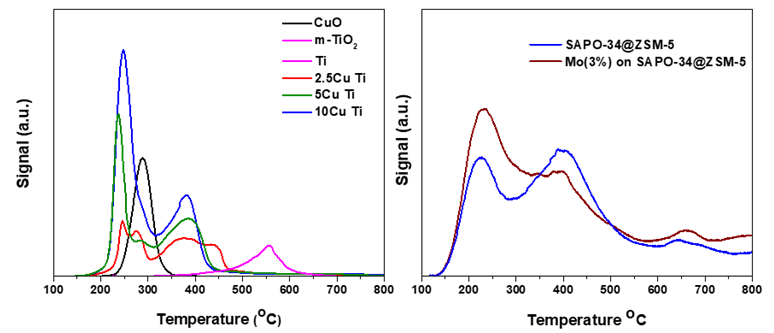
Temperature Programmed Reduction/Desorption /Oxidation (TPR/TPD/TPO) can be used to investigate desorption, oxidation and reduction as a function of the temperature; can be applied to characterize catalysts, materials related to Hydrogen and Fuel Cells, to identify the surface acidity/basicity as well as the dispersion of metals onto substrates.
X-ray analysis View all
SAXS Small Angle X-ray Scattering
SAXS is a non-destructive and versatile method to study the nanoscale structure of any type of material (solid, liquid, aerosols) ranging from new nanocomposites to biological macromolecules. Averaged particle sizes, shapes and distributions, porosity, degree of crystallinity and electron density maps with nanometer precision can be obtained.
XRI X-Ray Imaging
NanoCT with in situ nanomechanical tests (high mechanical stability and a maximum force of 0.8 N) enables the non-destructive structural study under load, of the inner structure of 3D samples. Resolution down to 50 nm. It can be adjusted for absorption and phase contrast and can be operated with a large field of view or at high resolution.
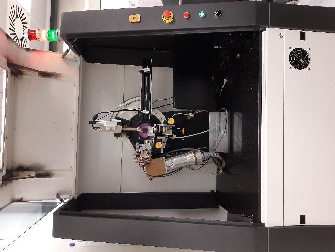
XRT X-Ray Tomography
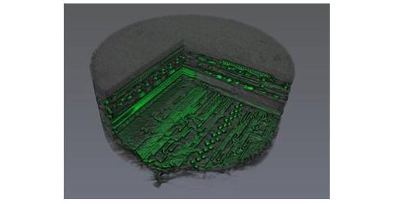
X-ray Computed Tomography (CT) is an indirect non-destructive technique for visualizing interior features within solid objects, and for obtaining digital information on their 3-D geometries and properties. Scanning x-ray tomography can also be performed using an x-ray beam (which size determines the resolution) focused on the sample.
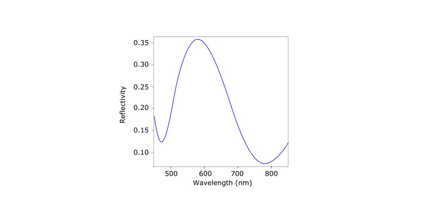
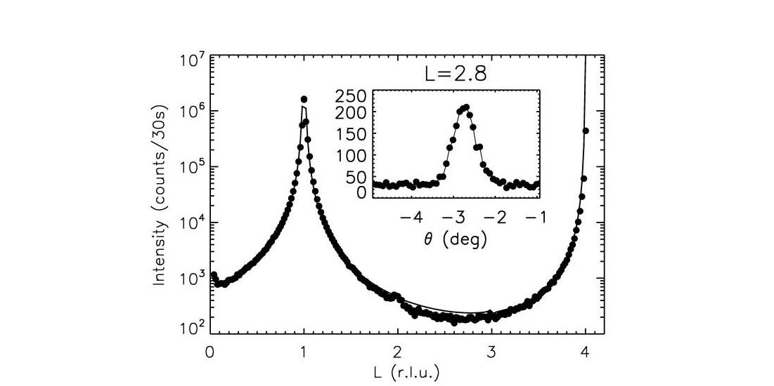
XRR X-Ray Reflectivity
X-Ray (specular) Reflectivity or X-Ray Reflectometry, XRR, is a grazing incidence X-ray elastic scattering technique sensitive to surface electron densities. It is used to characterize flat and relatively smooth surfaces and layered samples proofing information about superficial layers: thickness, density, roughness, index of refraction.
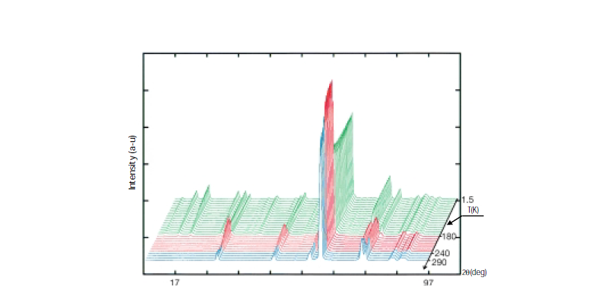
XRD X-Ray Diffraction
XRD provides non-destructive information on the structural order of a material. At large scattering angles XRD permits to identify different crystal phases and to quantify lattice distances and crystalline volume fractions. At low angles of incidence the surface roughness of a single crystal and the thickness of a deposition layer can be obtained.
XRM X-Ray Microscopy
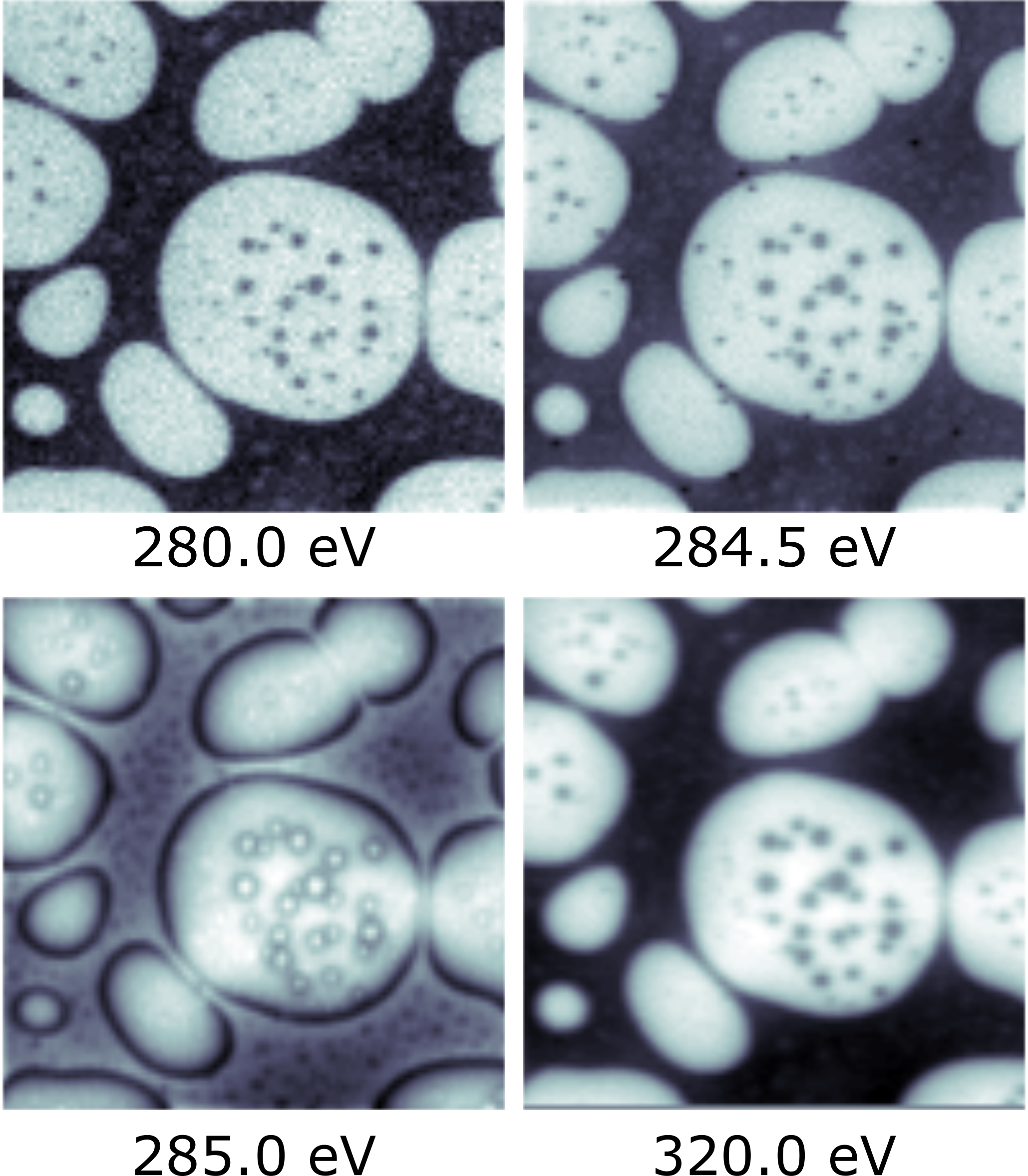
Scanning Transmission X-Ray Microscopy (STXM) is a non-invasive x-ray microscopy technique that enables the acquisition of images with elemental, chemical, and magnetic sensitivity. Typical STXM images exhibit a spatial resolution on the order of 10-20 nm, depending on the zone plate employed to focus the x-rays, and a typical lateral image size

